. 2020 Dec;34(12):15946-15960.
doi: 10.1096/fj.202001808R. Epub 2020 Oct 4.

Predicting susceptibility to SARS-CoV-2 infection based on structural differences in ACE2 across species

Affiliations

Predicting susceptibility to SARS-CoV-2 infection based on structural differences in ACE2 across species

Matthew R Alexander et al. FASEB J. 2020 Dec.

Abstract

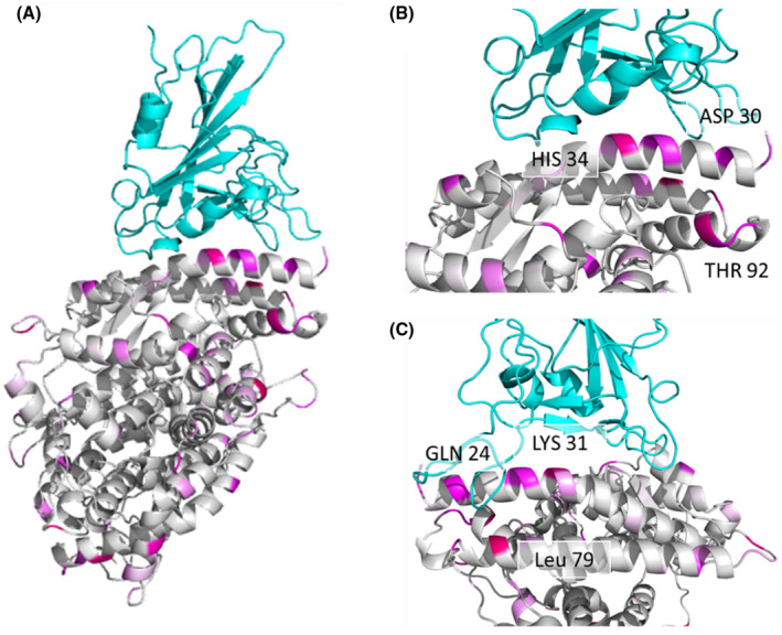
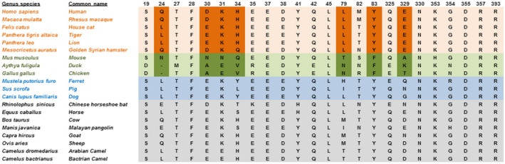
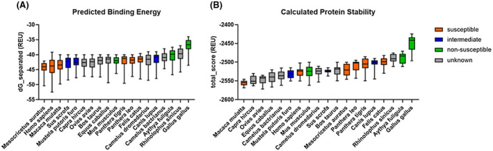
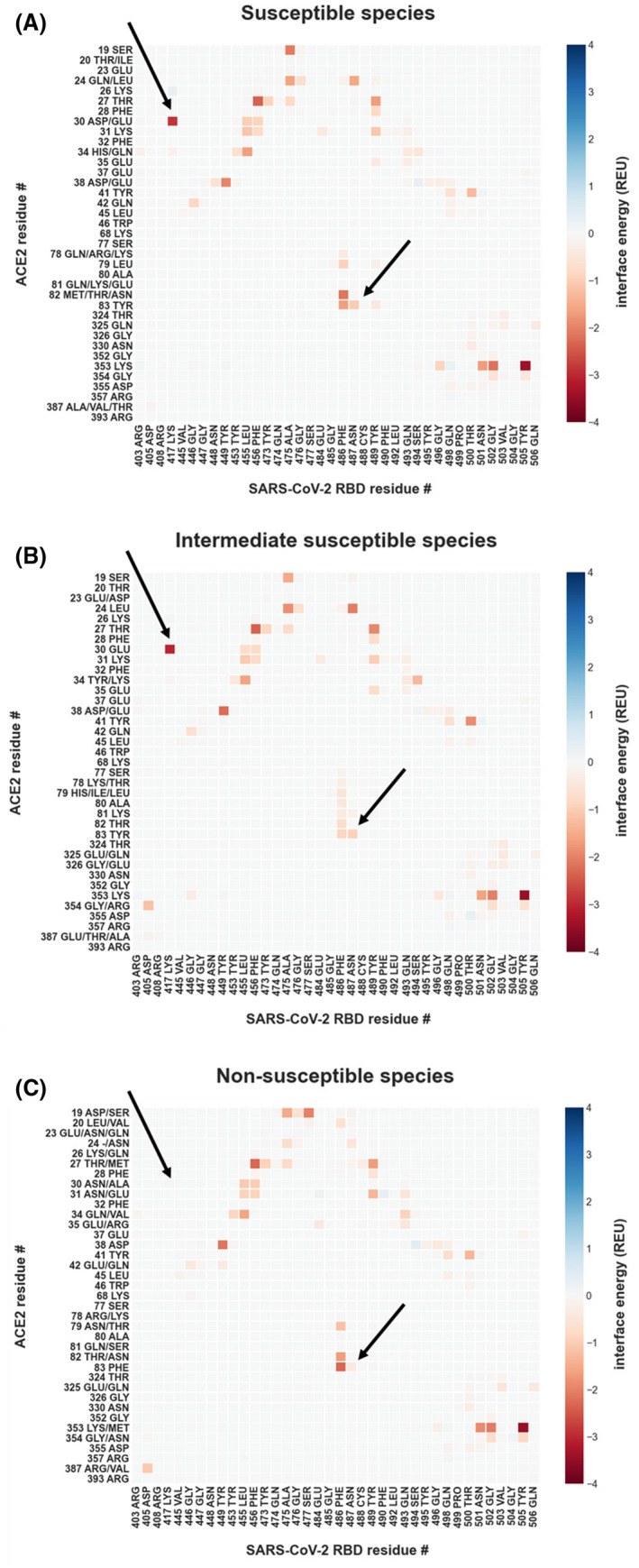
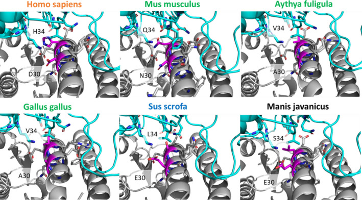
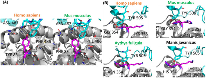
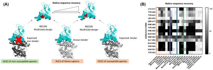
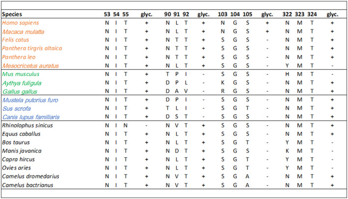
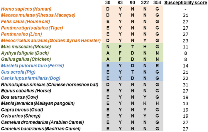
Severe acute respiratory syndrome coronavirus 2 (SARS-CoV-2) is the cause of the global pandemic of coronavirus disease-2019 (COVID-19). SARS-CoV-2 is a zoonotic disease, but little is known about variations in species susceptibility that could identify potential reservoir species, animal models, and the risk to pets, wildlife, and livestock. Certain species, such as domestic cats and tigers, are susceptible to SARS-CoV-2 infection, while other species such as mice and chickens are not. Most animal species, including those in close contact with humans, have unknown susceptibility. Hence, methods to predict the infection risk of animal species are urgently needed. SARS-CoV-2 spike protein binding to angiotensin-converting enzyme 2 (ACE2) is critical for viral cell entry and infection. Here we integrate species differences in susceptibility with multiple in-depth structural analyses to identify key ACE2 amino acid positions including 30, 83, 90, 322, and 354 that distinguish susceptible from resistant species. Using differences in these residues across species, we developed a susceptibility score that predicts an elevated risk of SARS-CoV-2 infection for multiple species including horses and camels. We also demonstrate that SARS-CoV-2 is nearly optimal for binding ACE2 of humans compared to other animals, which may underlie the highly contagious transmissibility of this virus among humans. Taken together, our findings define potential ACE2 and SARS-CoV-2 residues for therapeutic targeting and identification of animal species on which to focus research and protection measures for environmental and public health.

Keywords: COVID-19; angiotensin-converting enzyme 2; protein structural elements; severe acute respiratory syndrome coronavirus 2.


Conflict of interest statement

The authors declare that there are no conflicts of interest in connection with this article.

Figures

FIGURE 1
FIGURE 1
Multiple residues with high GroupSim scores are present at the interaction interface of the SARS‐CoV‐2 RBD and ACE2 complex. (A) SARS‐CoV‐2 RBD (top) and human ACE2 (bottom) complex shown as a ribbon diagram with GroupSim scores color coded in magenta. Higher scores are brighter in color. (B) Close‐up view of the interface highlighting ACE2 residues with high GroupSim scores. (C) Close‐up view after 90‐degree rotation from (B) demonstrating additional residues at the interface with high GroupSim scores
FIGURE 2
FIGURE 2
Twenty‐four key residues for SARS‐CoV‐2 RBD and ACE2 interactions. Highlighted residues are most similar in susceptible and different in non‐susceptible species as determined by GroupSim (Table S1). Susceptible species are in orange, non‐susceptible in green, intermediate in blue, and unknown in black/grey. Letters indicate amino acids using single‐letter naming
FIGURE 3
FIGURE 3
SARS‐CoV‐2 RBD has lower predicted binding energy and protein complex stability for ACE2 from non‐susceptible avian species. (A) Predicted binding energy as calculated with Rosetta and (B) protein complex stability of SARS‐CoV‐2 RBD and ACE2 of various species predicted by Rosetta
FIGURE 4
FIGURE 4
Energetic modeling of residue‐residue interactions identifies a link between ACE2 D30 and Y83 and SARS‐CoV‐2 susceptibility. Residue‐residue interactions are calculated with Rosetta, using the co‐crystal structure of the human ACE2 in complex with the SARS‐CoV‐2‐RBD (PDB: 6LZG and 6M0J) after backbone‐constrained relaxation for all interactions greater than 0.05 Rosetta Energy Units (REU) or smaller than −0.05 REU. Interactions are presented as the mean for all included samples. Residues depicted on the y‐axis are all observed amino acid identities for the particular position in its susceptibility group. (A) Per‐residue interactions for (A) susceptible species (human, cat, lion, tiger, hamster, and rhesus macaque), (B) intermediate susceptibility species (pig, dog, and ferret), and (C) non‐susceptible species (duck, mouse, and chicken). The arrows point to interactions that are not observed in non‐susceptible species
FIGURE 5
FIGURE 5
Binding interactions of ACE2 position 30 differ across species. Close‐up of the differences in binding interactions of positions 30 and 34 (magenta) of ACE2 from each species with the SARS‐CoV‐2 RBD. Position 30 is occupied by aspartic acid (D) in susceptible humans (Homo sapiens), is an asparagine (N) in non‐susceptible mice (Mus musculus), and an alanine (A) in the avian species (Aythya fuligula and Gallus gallus). Glutamic acid (E) is present at position 30 in pig (Sus scrofa) and Malayan pangolin (Manis javanica), representing intermediate and unknown susceptible species, respectively. Position 34 is conserved as histidine (H) in all susceptible species such as humans, yet has another residue identity in intermediate and non‐susceptible species. Species names in orange are susceptible, green are non‐susceptible, blue are intermediate susceptibility, and black are unknown
FIGURE 6
FIGURE 6
Binding interactions of ACE2 positions 83 and 354 differ across susceptible and non‐susceptible species. (A) ACE2 position 83 (magenta) is a tyrosine in the human susceptible species (left) and phenylalanine in the non‐susceptible mouse species (right). Tyrosine 83 of human ACE2 interacts with asparagine 87 of SARS‐CoV‐2 RBD, probably via a hydrogen bond. Phenylalanine in mouse ACE2 cannot interact with asparagine 487 due to the lack of a hydrogen bond donor. (B) Interactions of tyrosine r505 of the SARS‐CoV‐2‐RBD (cyan) with ACE2 residues 353 and residue 354 (magenta). ACE2 residue 353 is conserved as lysine with the only exception of histidine in the mouse ACE2. ACE2 residue 354 is glycine in the susceptible species (human), but an asparagine in non‐susceptible duck and chicken, and histidine in pangolin (unknown susceptibility). Species names in orange are susceptible, green are non‐susceptible, and black are unknown
FIGURE 7
FIGURE 7
Multistate design reveals SARS‐CoV‐2 RBD Tyr505 to have low native sequence recovery in non‐susceptible duck and chicken. (A) RECON multistate design overview. In the presence of ACE2 from two different species, the SARS‐CoV‐2‐RBD interface is redesigned. When two true binders are redesigned they should require few sequence changes, thus resulting in a higher native sequence recovery. In contrast, if the native sequence recovery for the interface residues is lower, then many sequence changes are required, indicating that one of the ACE2 proteins is a non‐binder. (B) Residue‐specific native sequence recovery as determined from RECON multistate design against the SARS‐CoV‐2‐RBD complex with human ACE2. Only residues of the SARS‐CoV‐2‐RBD, which are in the protein‐protein interface and show changes are depicted. Tyrosine 505 of SARS‐CoV‐2 RBD shows low native sequence recovery (black) in non‐susceptible duck (Gallus gallus) and chicken (Aythya fuligula). The orange box outlines susceptible species, the blue box outlines species with intermediate susceptibility, and the green box outlines non‐susceptible species
FIGURE 8
FIGURE 8
Predicted glycosylation profiles for ACE2 amino acid positions 53, 90, 103, and 322. Susceptible species are in orange, non‐susceptible in green, intermediate in blue, and unknown in black. + indicates presence,—indicates the absence of glycosylation. glyc = glycosylation. Letters indicate amino acids using single‐letter naming
FIGURE 9
FIGURE 9
Key residues of aligned ACE2 proteins with calculated SARS‐CoV‐2 susceptibility score for each species. Susceptible (orange), non‐susceptible (green), intermediate (blue), and unknown (black/grey) species are indicated

References

    1. Adhikari SP, Meng S, Wu YJ, et al. Epidemiology, causes, clinical manifestation and diagnosis, prevention and control of coronavirus disease (COVID‐19) during the early outbreak period: a scoping review. Infect Dis Poverty. 2020;9:29. - PMC - PubMed
    1. Zhang T, Wu Q, Zhang Z. Probable pangolin origin of SARS‐CoV‐2 associated with the COVID‐19 outbreak. CB. 2020;30:1346‐1351.e1342. - PMC - PubMed
    1. APHISpress@usda.gov . (April 6, 2020). USDA Statement on the Confirmation of COVID‐19 in a Tiger in New York. United States Department of Agriculture Animal and Plant Health Inspection Service. https://www.aphis.usda.gov/aphis/newsroom/news/sa_by_date/sa‐2020/ny‐zoo...
    1. APHISpress@usda.gov . (August 13, 2020). Confirmed cases of SARS‐CoV‐2 in animals in the United States. United States Department of Agriculture Animal and Plant Health Inspection Service. https://www.aphis.usda.gov/aphis/ourfocus/animalhealth/SA_One_Health/sar...
    1. Chan JF, Zhang AJ, Yuan S, et al. Simulation of the clinical and pathological manifestations of Coronavirus Disease 2019 (COVID‐19) in golden Syrian hamster model: implications for disease pathogenesis and transmissibility. Clin Infect Dis. 2020. 10.1093/cid/ciaa325 - DOI - PMC - PubMed

Publication types

Substances