. 2020 Dec;8(4):659-671.
doi: 10.1002/iid3.355. Epub 2020 Oct 4.

Disruption in the balance between apolipoprotein A-I and mast cell chymase in chronic hypersensitivity pneumonitis

Affiliations

Disruption in the balance between apolipoprotein A-I and mast cell chymase in chronic hypersensitivity pneumonitis

Yukihisa Inoue et al. Immun Inflamm Dis. 2020 Dec.

Abstract

Background: Apolipoprotein A-I (apoA-I) has an antifibrotic effect in idiopathic pulmonary fibrosis. Although pulmonary fibrosis is associated with poor prognosis of patients with hypersensitivity pneumonitis (HP), little is known regarding the role of apoA-I in the pathogenesis of HP.

Methods: Two-dimensional electrophoresis, immunoblotting, and enzyme-linked immunosorbent assays were performed for the identification and quantification of apoA-I in bronchoalveolar lavage fluid (BALF) from patients with acute and chronic HP. To investigate the degradation of apoA-I, apoA-I was incubated with BALF. Moreover, the role of apoA-I in TGF-β1-induced epithelial-mesenchymal transition of A549 cells was examined.

Results: The concentration of apoA-I in the BALF was significantly lower in chronic HP (n = 56) compared with acute HP (n = 31). The expression level of apoA-I was also low in the lung tissues of chronic HP. ApoA-I was degraded by BALF from HP patients. The number of chymase-positive mast cells in the alveolar parenchyma was inversely correlated with apoA-I levels in the BALF of chronic HP patients. In vitro experiment using A549 cells, untreated apoA-I inhibited TGF-β1-induced epithelial-mesenchymal transition, although this trend was not observed in the chymase-treated apoA-I.

Conclusions: A decrease of apoA-I was associated with the pathogenesis of chronic HP in terms of pulmonary fibrosis and mast cell chymase attenuated the protective effect of apoA-I against pulmonary fibrosis. Furthermore, apoA-I could be a crucial molecule associated with lung fibrogenesis of HP.

Keywords: apolipoprotein A-I; chymase; hypersensitivity pneumonitis; mast cell.


Conflict of interest statement

The authors declare that there are no conflict of interests.

Figures

Figure 1
Figure 1
(A) Images of two‐dimensional electrophoresis separation of pooled BALF proteins from patients with acute and chronic HP. Magnifying apoA‐I lesion, acute HP has spots with higher intensities. (B) Focusing on apoA‐I lesion, proteins are visualized with SYPRO‐Ruby and apoA‐I antibody by immunoblotting. The proteins indicated by the numbers were also identified as apoA‐I by using LC‐nESI‐MS/MS. Spot numbers were referred to the annotations in Table S1. apoA‐I, apolipoprotein A‐I; BALF, bronchoalveolar lavage fluid; HP, hypersensitivity pneumonitis
Figure 2
Figure 2
ELISA for measurement of apoA‐I concentrations in BALF and representative immunohistochemistry of apoA‐I proteins. (A) ApoA‐I concentrations in BALF from patients with HV, acute HP, and chronic HP. (B) ApoA‐I concentrations in BALF according to histological patterns of chronic HP. (C) Immunohistochemical staining of apoA‐I in lung tissues of control and chronic HP (UIP pattern) are shown. The expression of apoA‐I is lower in fibrotic lesions of chronic HP compared with normal lung tissues. Bars showed the mean values ± SEM. apoA‐I, apolipoprotein A‐I; BALF, bronchoalveolar lavage fluid; ELISA, enzyme‐linked immunosorbent assay; HP, hypersensitivity pneumonitis; HV, healthy volunteers; c‐NSIP, cellular nonspecific interstitial pneumonia; f‐NSIP, fibrotic nonspecific interstitial pneumonia; OP, organizing pneumonia; UIP, usual interstitial pneumonia; n.s., not significant. Scale bars = 500 µm and 50 µm in insets. ****p < .0001
Figure 3
Figure 3
Immunoblotting for apoA‐I in the mixture of lipid‐free apoA‐I and pooled BALF from HV and patients with acute and chronic HP. Fragmentation (arrow) and degradation (arrowhead) of apoA‐I were observed over a time‐course. The degradation of apoA‐I was inhibited by chymostatin and SBTI, but not by leupeptin, in acute and chronic HP. apoA‐I, apolipoprotein A‐I; BALF, bronchoalveolar lavage fluid; HP, hypersensitivity pneumonitis; HV, healthy volunteers; SBTI, soybean trypsin inhibitor
Figure 4
Figure 4
(A) The chymase concentration in BALF of HV, acute HP, and chronic HP. Acute HP, 36.7 ± 8.98 pg/ml; chronic HP, 9.51 ± 2.02 pg/ml; and HV, 2.59 ± 0.87 pg/ml. (B) Relationship between the concentration of chymase and the concentration of apoA‐I in BALF from patients with acute and chronic HP. apoA‐I, apolipoprotein A‐I; BALF, bronchoalveolar lavage fluid; HP, hypersensitivity pneumonitis; HV, healthy volunteers. Bars showed the mean ± SEM. **p < .01, ****p < .0001
Figure 5
Figure 5
Tryptase‐positive mast cells (MCT) and chymase‐positive mast cells (MCTC) in the BALF and lungs. (A) Representative images of immunohistochemical double staining for MCT (permanent red) and MCTC (DAB, brown). (B) The percentage of total mast cells and (C) MCTC in BALF from patients with acute HP, chronic HP, and HV. (D–F) Representative images of MCT and MCTC in alveolar parenchyma from normal lung tissues, acute HP, and chronic HP (UIP‐like lesion) are shown. (G) Total mast cell density per mm2 and (H) the proportion of MCTC in alveolar parenchyma from normal lung and fibrotic alveolar parenchyma from chronic HP. (I) Relationship between MCTC density in alveolar parenchyma and apoA‐I concentrations in BALF in chronic HP (Spearman's correlation coefficient r = −.52; p = .005). apoA‐I, apolipoprotein A‐I; BALF, bronchoalveolar lavage fluid; DAB, 3,3′‐diaminobenzidine; HP, hypersensitivity pneumonitis; HV, healthy volunteers; UIP, usual interstitial pneumonia. Bars showed the mean ± SEM. Scale bars = 500 µm and 50 µm in insets. *p < .05, **p < .01, ***p < .001, ****p < .0001
Figure 6
Figure 6
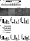
Inhibition of TGF‐β1‐induced EMT in A549 cells through the degradation of apoA‐I. (A) Immunoblotting of apoA‐I (1 mg/ml) in the absence or presence of chymase (0.001 U/ml BTEE) for the indicated times. (B) Untreated apoA‐I inhibits TGF‐β1‐induced morphological changes, but not chymase‐treated apoA‐I. The expression changes of (C) mRNA and (D) protein in EMT‐related markers, such as E‐cadherin, N‐cadherin, and vimentin, following TGF‐β1‐induced EMT in untreated apoA‐I or chymase‐treated apoA‐I. apoA‐I, apolipoprotein A‐I; ct‐ApoA‐I, chymase‐treated apoA‐I; BTEE, N‐benzoyl‐l‐tyrosine ethyl ester; EMT, epithelial–mesenchymal transition; mRNA, messenger RNA; TGF‐β1, transforming growth factor β. Six independent experiments were performed. Data are represented as the mean ± SEM. *p < .05, **p < .01 (untreated apoA‐I vs. TGF‐β1), # p < .05 (chymase‐treated apoA‐I vs. untreated apoA‐I)

Similar articles

Cited by

References

    1. Fink JN. Hypersensitivity pneumonitis. J Allergy Clin Immunol. 1984;74:1‐10. - PubMed
    1. Inoue Y, Ishizuka M, Furusawa H, et al. Acute inflammatory and immunologic responses against antigen in chronic bird‐related hypersensitivity pneumonitis. Allergol Int. 2019;68:321‐328. - PubMed
    1. Lacasse Y, Selman M, Costabel U, et al. Classification of hypersensitivity pneumonitis: a hypothesis. Int Arch Allergy Immunol. 2009;149:161‐166. - PubMed
    1. Vourlekis JS, Schwarz MI, Cherniack RM, et al. The effect of pulmonary fibrosis on survival in patients with hypersensitivity pneumonitis. Am J Med. 2004;116:662‐668. - PubMed
    1. Ohtani Y, Saiki S, Kitaichi M, et al. Chronic bird fancier's lung: histopathological and clinical correlation. An application of the 2002 ATS/ERS consensus classification of the idiopathic interstitial pneumonias. Thorax. 2005;60:665‐671. - PMC - PubMed