. 2020 Dec;111(12):4417-4428.
doi: 10.1111/cas.14676. Epub 2020 Oct 27.

β2-AR activation promotes cleavage and nuclear translocation of Her2 and metastatic potential of cancer cells

Affiliations

β2-AR activation promotes cleavage and nuclear translocation of Her2 and metastatic potential of cancer cells

Dan Liu et al. Cancer Sci. 2020 Dec.

Abstract

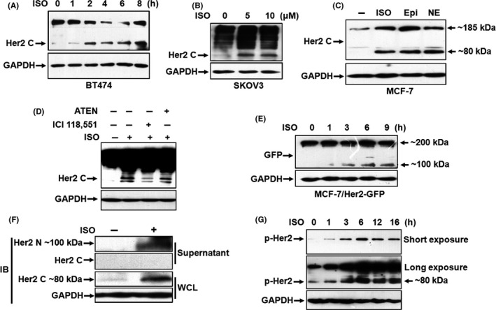
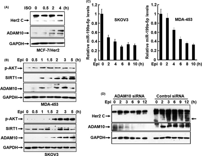
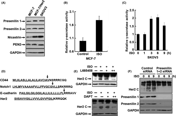
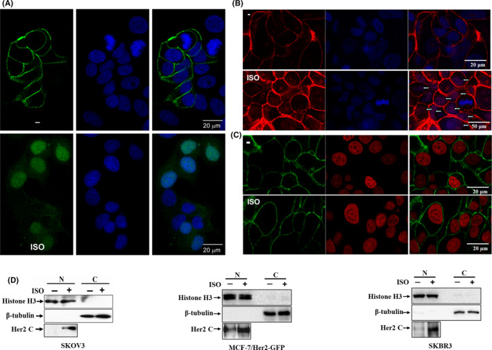
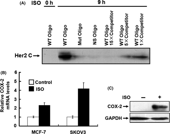
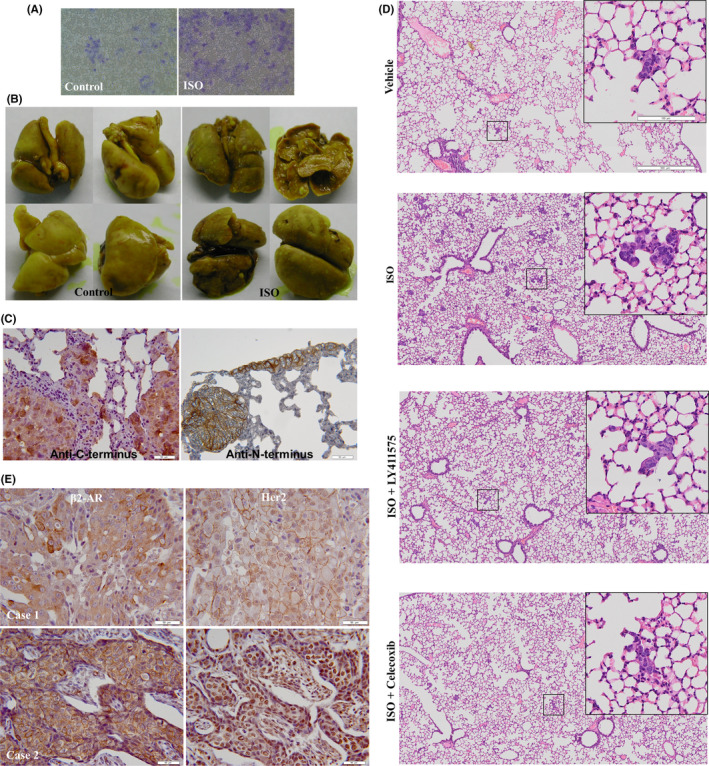
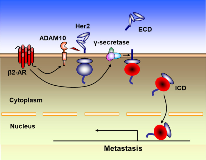
Prolonged hypersecretion of catecholamine induced by chronic stress may correlate with malignant progression of cancer. β2-adrenergic receptor (β2-AR) overexpressed in certain cancer cells may translate the signals from neuroendocrine system to malignant signals by interacting with oncoproteins, such as Her2. In the present study, we demonstrate that catecholamine stimulation activates the expression and proteolytic activity of ADAM10 by modulating the expression of miR-199a-5p and SIRT1 and also confirm that catecholamine induction triggers the activities of γ-secretase, leading to shedding of Her2 extracellular domain (ECD) by ADAM10 and subsequent intramembranous cleavage of Her2 intracellular domain (ICD) by presenilin-dependent γ-secretase, nuclear translocation of Her2 ICD, and enhanced transcription of tumor metastasis-associated gene COX-2. Chronic stimulation of catecholamine strongly promotes the invasive activities of cancer cells in vitro and spontaneous tumor lung metastasis in mice. Furthermore, nuclear localization of Her2 was significantly correlated with overexpression of β2-AR in human breast cancer tissues, indicating that catecholamine-induced β2-AR activation plays decisive roles in tumor metastasis. Our data also reveal that an unknown mechanism by which the regulated intramembrane proteolysis (RIP) initiated by β2-AR-mediated signaling controls a novel Her2-mediated signaling transduction.

Keywords: Her2; a disintegrin and metalloprotease 10; metastasis; β2-adrenergic receptor; γ-secretase.


Conflict of interest statement

The authors declare that there are no potential conflicts of interest.

Figures

FIGURE 1
FIGURE 1
Catecholamine stimulation induces cleavage and phosphorylation of Her2. A, BT474 cells were starved overnight and then treated with 5 μmol/L isoproterenol (ISO) for 0, 1, 2, 4, 6 or 8 h. Expression of Her2 was analyzed by western blot with the antibodies against the C‐terminus of Her2. Equal loading was verified by detection of GAPDH. B, SKOV3 cells were treated with 0, 5 or 10 μmol/L ISO for 6 h. C, MCF‐7 cells were treated with 5 μmol/L ISO, 10 μmol/L epinephrine (Epi) or 10 μmol/L norepinephrine (NE). The arrows on the right indicate molecular weight of the bands. D, MCF‐7 cells were pretreated with 1 μmol/L ICI‐118551 or 1 μmol/L ATEN for 1 h and then incubated with 5 μmol/L ISO. E, MCF‐7/Her2‐GFP cells were treated with 5 μmol/L ISO for 0, 1, 3, 6 or 9 h. Western blot was performed by using the antibody against GFP. The arrows on the right indicate molecular weight of the bands. F, The supernatant and whole‐cell lysates of SKOV3 cells treated with 5 μmol/L ISO were collected and the fragments of Her2 detected by the antibodies against the N‐terminus and C‐terminus of Her2. G, SKOV3 cells were treated with ISO for 0, 1, 2, 6, 12 or 16 h. Phosphorylation of Her2 was detected by western blot
FIGURE 2
FIGURE 2
Catecholamine induces the cleavage of Her2 ECD by promoting ADAM10 expression. A, MCF‐7/Her2 cells were treated with 5 μmol/L isoproterenol (ISO) for 0, 0.5, 2 or 4 h. Expression of Her2 and ADAM10 was analyzed by western blot with the antibodies against the C‐terminus of Her2 and ADAM10. B, MDA‐453 and SKOV3 cells were treated with 10 μmol/L of epinephrine (Epi) for 0, 0.5, 1, 1.5, 2, 3, 5 or 7 h. Phosphorylated Akt, SIRT1, and ADAM10 were analyzed by western blot. C, SKOV3 and MDA453 cells were treated with 10 μmol/L of epinephrine for 0, 2, 4, 6, 8 or 10 h. The expression of miR‐199a‐5p was analyzed by real‐time PCR. D, SKOV3 cells were transfected with the specific siRNA targeting ADAM10 or control siRNA. After transfection for 48 h, the cells were treated with 10 μmol/L of epinephrine for 0, 2, 3, 6, 9 or 12 h and the expression of Her2 and ADAM10 was detected by western blot with the antibodies against the C‐terminus of Her2 and ADAM10. The arrow on the right indicates molecular weight of the bands
FIGURE 3
FIGURE 3
γ‐Secretase activity induced by catecholamine stimulation is responsible for the generation of p80Her2 intracellular domain (ICD). A, Expression of presenilin 1, presenilin 2, nicastrin, and PEN‐2 in MCF‐7, MCF‐7/Her2 and SKOV3 cells was analyzed by western blot. B, Cell lysates from MCF‐7 cells treated with 5 μmol/L ISO were prepared and γ‐secretase activities measured. C, SKOV3 cells were treated with 5 μmol/L of ISO for 0, 1, 3, 6 or 9 h. The activities of γ‐secretase were measured. D, Comparison of the known γ‐secretase substrates CD44, Notch and E‐cadherin with Her2 for homology of the amino acid sequences of the transmembrane domain (underlined sequences). The arrows indicated the identified cleavage sites. E, SKOV3 cells were pretreated with 0.5 and 2 μmol/L L685458 or 1 and 5 μmol/L DAPT and then incubated with 5 μmol/L ISO. Expression of Her2 was analyzed by western blot. F, SKOV3 cells were transfected with siRNA targeting presenilin 1 and presenilin 2. After transfection for 48 h, the cells were treated with 5 μmol/L isoproterenol (ISO) for 0, 6 or 9 h and the expression of Her2, presenilin 1, and presenilin 2 was detected by western blot
FIGURE 4
FIGURE 4
Catecholamine stimulation mediates nuclear translocation of Her2 intracellular domain (ICD). A, MCF‐7/Her2‐GFP cells were treated with 5 μmol/L of isoproterenol (ISO). The subcellular localization of Her2‐GFP fusion protein was observed under a confocal microscope. The nuclei were stained with 1 μg/mL DAPI. Bar = 20 μm B, SKBR3 cells were labeled with the antibody against the C‐terminus of Her2. Binding was detected with Alexa fluor 549 (Red)‐labeled secondary antibody. The arrows indicate the nuclei staining. Nuclei were also stained with DAPI. Bar = 20 μm (upper panel) or 50 μm (lower panel). C, SKBR3 cells were incubated with the antibody against N‐terminus of Her2 and binding was detected with Alexa fluor 488 (green)‐labeled secondary antibody. Nuclei were stained with propidium iodide (PI). Bar = 20 μm. D, SKOV3, MCF‐7/Her2 and SKOV3 cells were treated with 5 μmol/L of ISO for 0 or 9 h after overnight starvation. The nuclear extracts were prepared using a Nuclear‐Cytosol Extraction Kit. The expression of Her2 was analyzed by the antibody against the C‐terminus of Her2. Detection of histone H3 and β‐tubulin was used as the indicators of nuclear and cytoplasmic proteins
FIGURE 5
FIGURE 5
Her2 intracellular domain (ICD) physically binds to the promoter of COX2 gene and drives transactivation of COX2 gene. A, 200 μg of the nuclear extracts was incubated with the 5′‐biotinylated double‐stranded oligonucleotides (5′‐ATAAACTTCAAATTTCAGTA‐3′) corresponding to the positions −1770 to −1750 of the COX‐2 promoter previously coupled to Dynabeads M‐280. The protein/DNA complexes were separated with a Dynal magnet, denatured in SDS sample buffer and subjected to SDS‐PAGE. Binding of Her2 was detected by western blot with the antibodies against N‐terminus or C‐terminus of Her2. The same double‐stranded sequences that are not biotinylated were used as the competitors. The biotinylated oligonucleotides containing the mutated Her2 binding sequences (5′‐ATAAACTGACCCGGGAAGTA‐3′) and the sequences lacking the Her2 binding motif (5′‐ATAAACTTCAAATTTCAGTA‐3′) were used as the controls. B, C, MCF‐7 and SKOV3 cells were treated with 5 μmol/L of isoproterenol (ISO). The expression of COX‐2 at the mRNA and protein levels was detected by real‐time RT‐PCR (B) and western blot (C)
FIGURE 6
FIGURE 6
Catecholamine stimulation promotes the invasive and metastatic potentials of tumor cells. A, SKOV3 cells were chronically stimulated by 5 μmol/L isoproterenol (ISO) and invasive activities of the cells assessed by a cell invasion assay kit. The invasive cells were fixed and stained. B, A total of 0.1 mL of the cell suspension (108 cells/mL) was injected sc in the right upper flank of 5‐ to 6‐wk‐old athymic female BALB/c nude mice. After tumor cell injection for 4 d, the mice were treated daily ip with PBS or ISO (10 mg/kg). Eight mice were used in each group. At 60 d following tumor implantation, mice were sacrificed. The lungs of the mice were autopsied, fixed, and photographed. C, Lung metastatic tumors were dissected. Paraffin‐embedded tissue sections were stained with rabbit monoclonal antibodies against N‐terminus and C‐terminus of Her2. D, SKOV3 cells (8 × 105/mouse) were injected intravenously into NCG mice via the tail vein, and then the mice were treated daily with ISO (10 mg/kg, ip), celecoxib (COX‐2 inhibitor, 5 mg/kg, ip) or LY411,575 (γ‐secretase inhibitor, 1 mg/kg, po). Each group contained 5 mice. At 2 wk later, the mice were sacrificed and H&E staining was performed in the dissected lung tissue. E, Expression of β2‐AR and Her2 in breast cancer tissues was analyzed by immunohistochemistry with the antibodies against β2‐AR and C‐terminus of Her2. Bar = 50 μm
FIGURE 7
FIGURE 7
Molecular mechanism of cleavage and nuclear translocation of Her2. Catecholamine‐induced β2‐AR activation triggers shedding of Her2 extracellular domain (ECD) by ADAM10 and subsequent intramembranous cleavage of Her2 intracellular domain (ICD) by presenilin‐dependent γ‐secretase, resulting in nuclear translocation of p80Her2 and enhanced transcription of target genes

Similar articles

Cited by

References

    1. Thaker PH, Han LY, Kamat AA, et al. Chronic stress promotes tumor growth and angiogenesis in a mouse model of ovarian carcinoma. Nat Med. 2006;12:939‐944. - PubMed
    1. Sood AK, Armaiz‐Pena GN, Halder J, et al. Adrenergic modulation of focal adhesion kinase protects human ovarian cancer cells from anoikis. J Clin Invest. 2010;120:1515‐1523. - PMC - PubMed
    1. Cui B, Luo Y, Tian P, et al. Stress‐induced epinephrine enhances lactate dehydrogenase A and promotes breast cancer stem‐like cells. J Clin Invest. 2019;129:1030‐1046. - PMC - PubMed
    1. Noma T, Lemaire A, Naga Prasad SV, et al. Beta‐arrestin‐mediated beta1‐adrenergic receptor transactivation of the EGFR confers cardioprotection. J Clin Invest. 2007;117:2445‐2458. - PMC - PubMed
    1. Engelhardt S. Alternative signaling: cardiomyocyte beta1‐adrenergic receptors signal through EGFRs. J Clin Invest. 2007;117:2396‐2398. - PMC - PubMed

MeSH terms