. 2020 Nov 2;130(11):5847-5857.
doi: 10.1172/JCI138099.

HIV-1 viremia not suppressible by antiretroviral therapy can originate from large T cell clones producing infectious virus

Affiliations

HIV-1 viremia not suppressible by antiretroviral therapy can originate from large T cell clones producing infectious virus

Elias K Halvas et al. J Clin Invest. .

Abstract

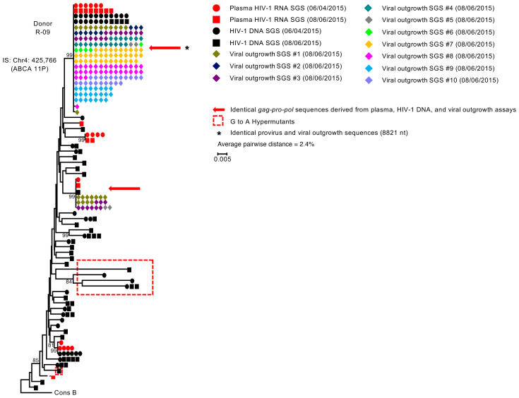
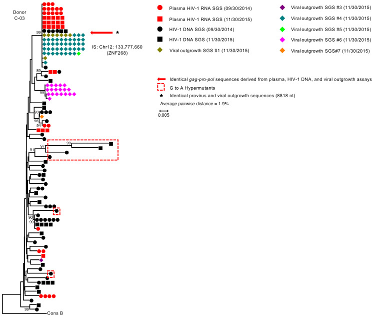
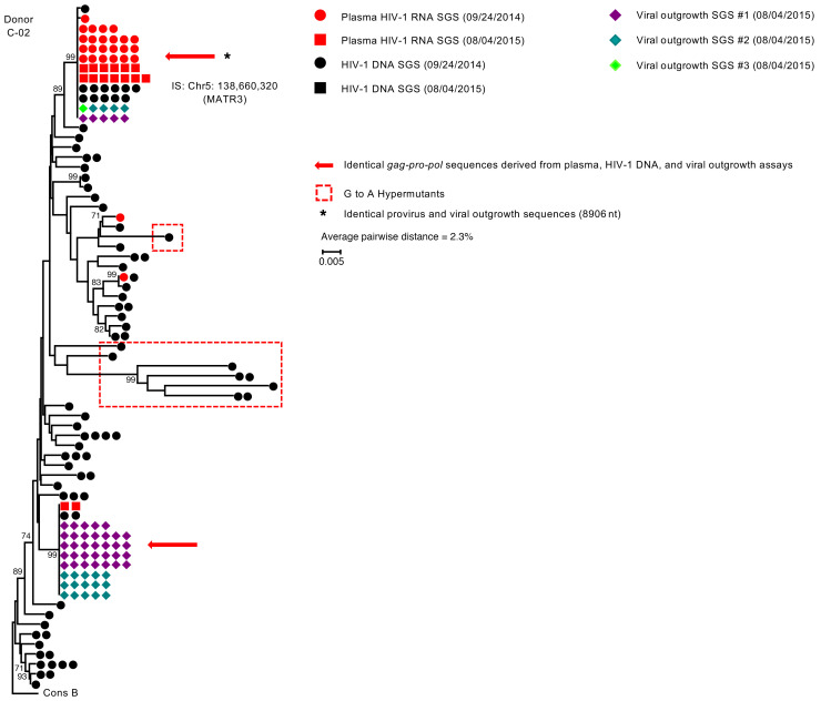
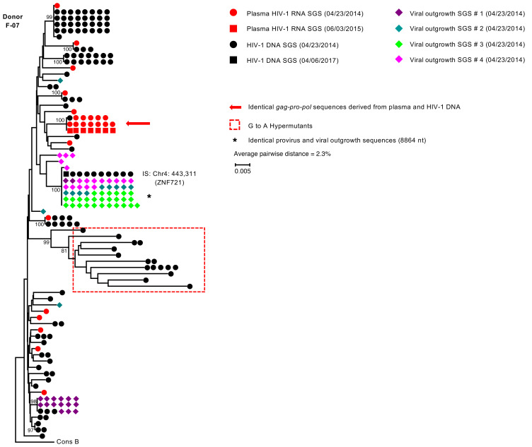
BACKGROUNDHIV-1 viremia that is not suppressed by combination antiretroviral therapy (ART) is generally attributed to incomplete medication adherence and/or drug resistance. We evaluated individuals referred by clinicians for nonsuppressible viremia (plasma HIV-1 RNA above 40 copies/mL) despite reported adherence to ART and the absence of drug resistance to the current ART regimen.METHODSSamples were collected from at least 2 time points from 8 donors who had nonsuppressible viremia for more than 6 months. Single templates of HIV-1 RNA obtained from plasma and viral outgrowth of cultured cells and from proviral DNA were amplified by PCR and sequenced for evidence of clones of cells that produced infectious viruses. Clones were confirmed by host-proviral integration site analysis.RESULTSHIV-1 genomic RNA with identical sequences were identified in plasma samples from all 8 donors. The identical viral RNA sequences did not change over time and did not evolve resistance to the ART regimen. In 4 of the donors, viral RNA sequences obtained from plasma matched those sequences from viral outgrowth cultures, indicating that the viruses were replication competent. Integration sites for infectious proviruses from those 4 donors were mapped to the introns of the MATR3, ZNF268, ZNF721/ABCA11P, and ABCA11P genes. The sizes of the clones were estimated to be from 50 million to 350 million cells.CONCLUSIONThese findings show that clones of HIV-1-infected cells producing virus can cause failure of ART to suppress viremia. The mechanisms involved in clonal expansion and persistence need to be defined to effectively target viremia and the HIV-1 reservoir.FUNDINGNational Cancer Institute, NIH; Howard Hughes Medical Research Fellows Program, Howard Hughes Medical Institute; Bill and Melinda Gates Foundation; Office of AIDS Research; American Cancer Society; National Cancer Institute through a Leidos subcontract; National Institute for Allergy and Infectious Diseases, NIH, to the I4C Martin Delaney Collaboratory; University of Rochester Center for AIDS Research and University of Rochester HIV/AIDS Clinical Trials Unit.

Keywords: AIDS/HIV; Clonal selection; T cells; Virology.


Conflict of interest statement

Conflict of interest: JWM has served as a consultant to Gilead Sciences, Merck Co. Inc., Xi’an Yufan Biotechnologies, and Accelevir Diagnostics, and owns share options in Co-Crystal Pharmaceuticals, Inc and Abound Bio.

Figures

Figure 1
Figure 1. Representative neighbor-joining p-distance phylogenetic tree of plasma HIV-1 RNA–, HIV-1 DNA–, and quantitative viral outgrowth assay–derived sequences from donor R-09 with intact replication-competent proviruses producing nonsuppressible viremia.
The trees were rooted to a subtype B consensus sequence. Single-genome sequences (SGSs) of a portion of gag (p6), all of pro, and the portion of pol encoding the first 300 amino acids of reverse transcriptase (gag-pro-pol) were obtained from plasma HIV-1 RNA, HIV-1 DNA in peripheral blood mononuclear cells (PBMCs), and culture supernatants from p24+ qVOA wells for donor R-09 (35). Red circles and squares represent plasma-derived sequences from 2 time points. Black circles and squares represent HIV-1 DNA–derived sequences from 2 time points. Different-colored diamonds represent viral outgrowth assay–derived sequences from independent p24+ quantitative viral outgrowth assay (qVOA) wells. A red arrow shows identical gag-pro-pol sequences from plasma HIV-1 RNA–, HIV-1 DNA–, and p24+ qVOA HIV-1 RNA–derived sequences. The asterisk shows matching sequences of provirus (near-full-length HIV-1 DNA and host–to–full-length provirus–to–host amplicons) and near-full-length viral RNA sequences from p24+ wells. HIV-1 DNA sequences with G to A hypermutations are enclosed in red hashed boxes. The viral outgrowth sequence variants that differ by 1 to 2 nucleotides can be attributed to either ex vivo replication or errors introduced during cDNA synthesis. Average pairwise distances (APDs) calculated by MEGA v6.0 using HIV-1 DNA sequences excluding hypermutated sequences. IS indicates the integration site location of the repliclone in the host genome.
Figure 2
Figure 2. Representative neighbor-joining p-distance phylogenetic tree of plasma HIV-1 RNA–, HIV-1 DNA–, and quantitative viral outgrowth assay–derived sequences from donor C-03 with intact replication-competent proviruses producing nonsuppressible viremia.
The tree was rooted to a subtype B consensus sequence. Single-genome sequences (SGSs) of a portion of gag (p6), all of pro, and the portion of pol encoding the first 300 amino acids of reverse transcriptase (gag-pro-pol) were obtained from plasma HIV-1 RNA, HIV-1 DNA in PBMCs, and culture supernatants from p24+ qVOA wells for donor C-03 (35). Red circles and squares represent plasma-derived sequences from 2 time points. Black circles and squares represent HIV-1 DNA–derived sequences from 2 time points. Different-colored diamonds represent viral outgrowth assay–derived sequences from independent p24+ quantitative viral outgrowth assay (qVOA) wells. A red arrow shows identical gag-pro-pol sequences from plasma HIV-1 RNA–, HIV-1 DNA–, and p24+ qVOA HIV-1 RNA–derived sequences. The asterisk shows matching sequences of provirus (near-full-length HIV-1 DNA and host–to–full-length provirus–to–host amplicons) and near-full-length viral RNA sequences from p24+ wells. HIV-1 DNA sequences with G to A hypermutations are enclosed in red hashed boxes. The viral outgrowth sequence variants that differ by 1 to 2 nucleotides can be attributed to either ex vivo replication or errors introduced during cDNA synthesis. Average pairwise distances (APDs) calculated by MEGA v6.0 using HIV-1 DNA sequences excluding hypermutated sequences. IS indicates the integration site location of the repliclone in the host genome.
Figure 3
Figure 3. Representative neighbor-joining p-distance phylogenetic tree of plasma HIV-1 RNA–, HIV-1 DNA–, and quantitative viral outgrowth assay–derived sequences from donor C-02 with intact replication-competent proviruses producing nonsuppressible viremia.
The tree was rooted to a subtype B consensus sequence. Single-genome sequences (SGSs) of a portion of gag (p6), all of pro, and the portion of pol that encodes the first 300 amino acids of reverse transcriptase (gag-pro-pol) were obtained from plasma HIV-1 RNA, HIV-1 DNA from PBMCs, and culture supernatants from p24+ qVOA wells for donor C-02 (35). Red circles and squares represent plasma-derived sequences from 2 different time points. Black circles and squares represent HIV-1 DNA–derived sequences from 2 different time points. Different-colored diamonds represent viral outgrowth assay–derived sequences from independent p24+ wells. A red arrow shows identical gag-pro-pol sequences for plasma-, HIV-1 DNA–, and viral outgrowth assay HIV-1 RNA–derived sequences. The asterisk shows matching sequences of provirus (near-full-length HIV-1 DNA and host–to–full-length provirus–to–host amplicons) and near-full-length viral RNA sequences from p24+ wells. HIV-1 DNA sequences with G to A hypermutations are enclosed in red hashed boxes. The viral outgrowth sequence variants that differ by 1 to 2 nucleotides can be attributed to either ex vivo replication or errors introduced during cDNA synthesis. Average pairwise distances (APDs) calculated by MEGA v6.0 using HIV-1 DNA sequences and excluding hypermutated sequences. IS indicates the integration site location of the repliclone in the host genome.
Figure 4
Figure 4. Representative neighbor-joining p-distance phylogenetic tree of plasma HIV-1 RNA–, HIV-1 DNA–, and quantitative viral outgrowth assay–derived sequences for donor F-07 with an intact replication-competent provirus.
The tree was rooted to a subtype B consensus sequence. Single-genome sequences (SGSs) of a portion of gag (p6), all of pro, and the portion of pol encoding the first 300 amino acids of reverse transcriptase (gag-pro-pol) were obtained from plasma HIV-1 RNA, HIV-1 DNA from PBMCs, and culture supernatants from p24+ qVOA wells for donor F-07 (35). Red circles and squares represent plasma-derived sequences from 2 different time points. Black circles and squares represent HIV-1 DNA–derived sequences from 2 different time points. Different-colored diamonds represent viral outgrowth assay–derived sequences from independent p24+ wells. A red arrow shows identical gag-pro-pol sequences from plasma- and HIV-1 DNA–derived sequences. The asterisk shows matching sequences of provirus (near-full-length HIV-1 DNA and host–to–full-length provirus–to–host amplicons) and near-full-length viral RNA sequences from p24+ wells. HIV-1 DNA sequences with G to A hypermutations are enclosed in red hashed boxes. The viral outgrowth sequence variants that differ by 1–2 nucleotides can be attributed to either ex vivo replication or errors introduced during cDNA synthesis. Average pairwise distances (APDs) calculated by MEGA v6.0 using HIV-1 DNA sequences and excluding hypermutated sequences. IS indicates the integration site location of the repliclone in the host genome.

Comment in

References

    1. Palella FJ, et al. Declining morbidity and mortality among patients with advanced human immunodeficiency virus infection. HIV Outpatient Study Investigators. N Engl J Med. 1998;338(13):853–860. doi: 10.1056/NEJM199803263381301. - DOI - PubMed
    1. Finzi D, et al. Identification of a reservoir for HIV-1 in patients on highly active antiretroviral therapy. Science. 1997;278(14):1295–1300. - PubMed
    1. Wong JK, et al. Recovery of replication-competent HIV despite prolonged suppression of plasma viremia. Science. 1997;278(14):1291–1295. - PubMed
    1. Siliciano JD, et al. Long-term follow-up studies confirm the stability of the latent reservoir for HIV-1 in resting CD4+ T cells. Nat Med. 2003;9(6):727–728. doi: 10.1038/nm880. - DOI - PubMed
    1. Ho YC, et al. Replication-competent noninduced proviruses in the latent reservoir increase barrier to HIV-1 cure. Cell. 2013;155(3):540–551. doi: 10.1016/j.cell.2013.09.020. - DOI - PMC - PubMed

Publication types