

Review
doi: 10.1161/CIRCULATIONAHA.120.048186.
Epub 2020 Oct 5.
Progress Toward Cardiac Xenotransplantation
Affiliations
- PMID: 33017208
- PMCID: PMC7990044
- DOI: 10.1161/CIRCULATIONAHA.120.048186
Item in Clipboard
Review
Progress Toward Cardiac Xenotransplantation
Circulation.
.
Abstract
Consistent survival of life-supporting pig heart xenograft recipients beyond 90 days was recently reported using genetically modified pigs and a clinically applicable drug treatment regimen. If this remarkable achievement proves reproducible, published benchmarks for clinical translation of cardiac xenografts appear to be within reach. Key mechanistic insights are summarized here that informed recent pig design and therapeutic choices, which together appear likely to enable early clinical translation.
Keywords: cardiomyopathies; genetic engineering; heart failure; heart transplantation; heterografts; myocardial ischemia; transplantation, heterologous.
Figures
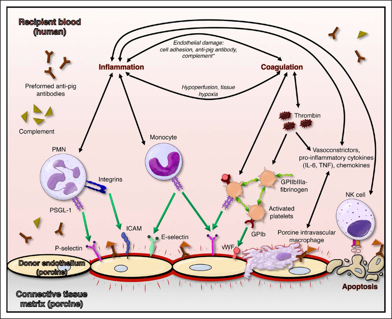
Preformed human anti-pig antibodies bind to porcine endothelium, triggering complement binding and Fc-receptor–mediated ligation of platelets and leukocytes and upregulation of adhesion molecules on both adherent formed blood elements and inflamed endothelium. Complement cascade activation (orange symbols), nonphysiological adhesion of human platelets to porcine endothelium, and absence of nonself signals (illustrated for natural killer cells) contribute to a prothrombotic, proinflammatory milieu that leads to loss of vascular barrier function and organ failure. GP indicates glycoprotein; ICAM, intercellular adhesion molecule; IL-6, interleukin-6; PMN, polymorphonuclear; PSGL-1, P-selectin glycoprotein ligand 1; TNF, tumor necrosis factor; and vWF, von Willebrand factor.
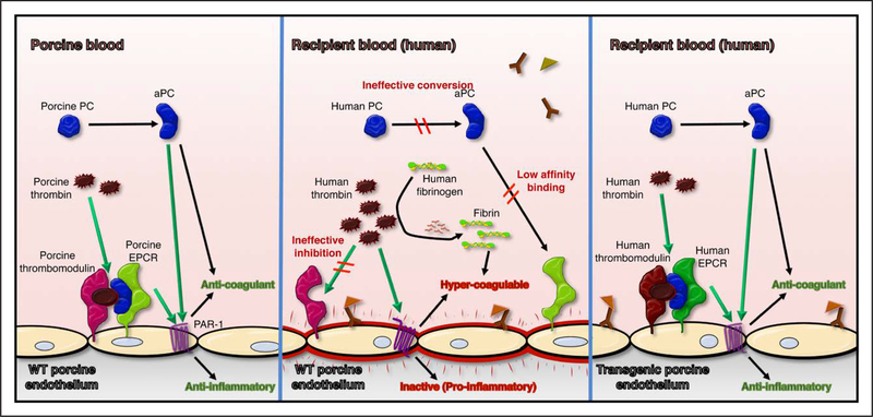
Relative to physiological regulation of thrombosis (left), porcine endothelium exposed to human blood is activated by binding of anti-pig antibodies, creating a prothrombotic environment (middle). Physiologically inappropriate amplification of blood clotting is contributed to by ineffective neutralization of human thrombin by porcine thrombomodulin, inefficient conversion of protein C (PC) to activated PC (aPC) by thrombin-thrombomodulin complex, and low-affinity binding of human aPC to porcine endothelial protein C receptor (EPCR), which in turn leads to inefficient thrombin degradation and reduced cytoprotective signaling through endothelial cell proteinase-activated receptor 1 (PAR-1). These molecular incompatibilities between species are addressed by expression of human thromboregulatory proteins, including human thrombomodulin and human EPCR (right), as well as human tissue factor pathway inhibitor (not illustrated). WT indicates wild-type.
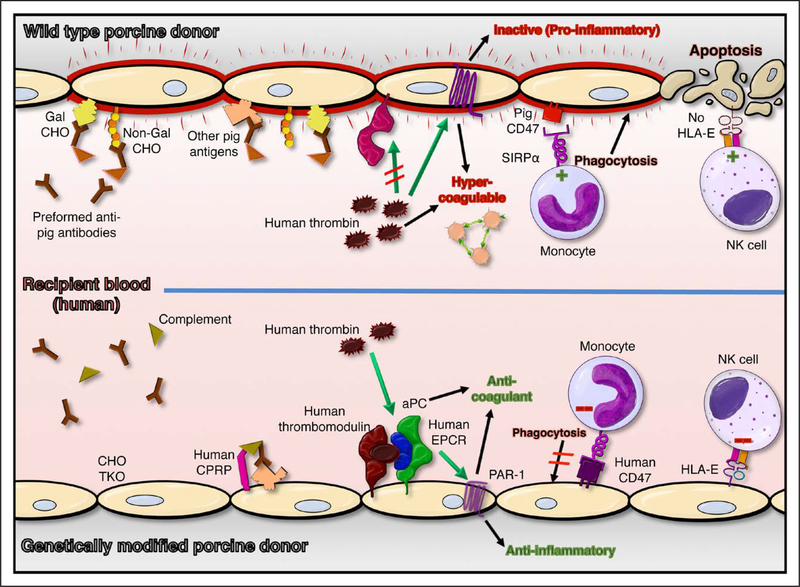
Examples of genetic modifications designed to prevent known xenograft injury (top) include Gal α1–3Gal (Gal) and 2 other carbohydrate (CHO) gene knockouts (TKO), and expression of human complement pathway regulatory proteins (hCPRPs), coagulation pathway regulatory proteins (eg, thrombomodulin [hTBM] and endothelial protein C receptor [hEPCR]), and self-recognition receptors (hCD47; human leukocyte antigen-E [HLA-E]; bottom). Absence of carbohydrate antigens (CHO TKO) and expression of human complement and coagulation pathway regulatory molecules reduce endothelial activation and injury and promote endothelial cytoprotective mechanisms. Expression of self-recognition receptors inhibits (red negative symbols, bottom) pathogenic mechanisms mediated by monocytes and natural killer (NK) cells that contribute to cross-species injury (green positive symbols, top). In addition to the pathways illustrated, hCD39, human tissue factor pathway inhibitor, hemoxygenase-1, and A20 are among the human genes included in some of the various multigene pig constructs that are currently under preclinical evaluation. aPC indicates activated protein C; PAR-1, proteinase-activated receptor 1; and SIRPα, signal regulatory protein-α.
References
Publication types
MeSH terms
Grants and funding
LinkOut - more resources
Full Text Sources
Other Literature Sources
Medical
