

Biodistribution of andrographolide to assess the interior-exterior relationship between the lung and intestine using microPET
- PMID: 33017514
- PMCID: PMC7606023
- DOI: 10.1111/1759-7714.13682
Biodistribution of andrographolide to assess the interior-exterior relationship between the lung and intestine using microPET
Abstract
Background: One classic traditional Chinese medicine theory is that the "lung and intestine are exterior-interiorly related"; however, this has not been confirmed experimentally. The aim of this study was to provide a biological basis for the theory by measuring the tissue distribution of andrographolide.
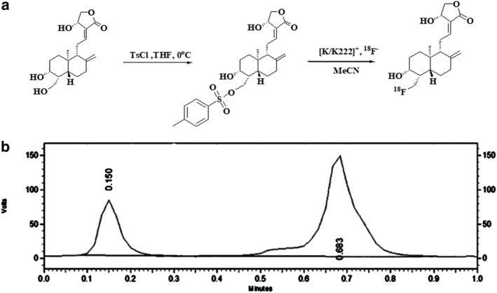
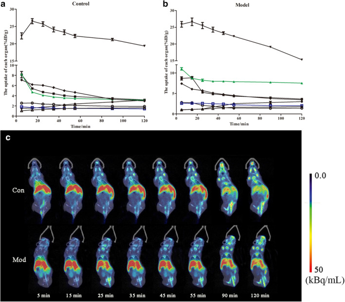
Methods: Acute pneumonia was induced in a mouse model by repeated stimulation with lipopolysaccharide. The distribution of andrographolide in mice was observed by positron emission tomography (PET) imaging with [18 F]-labeled andrographolide, and changes in the in vivo distribution before and after modeling were compared. Subsequently, the consistency of pathological changes in lung and intestine was confirmed by observation of pathological sections. Finally, the results were verified by cytokine detection.
Results: The value of organ uptake, pathological changes and inflammatory factor expression of the lung and intestine were consistent. The concentration of andrographolide in the lung and intestine increased significantly, and was confirmed by pathology and enzyme-linked immunosorbent assays (ELISA).
Conclusions: Micro-positron emission tomography (microPET) can be used to visually observe the distribution of medicinal ingredients in vivo, and [18 F]-andrographolide can be used as a tool to assess the interior-exterior relationship between the lung and intestine.
Keywords: Andrographolide; enteritidis; interior-exterior relationship; pneumonia; positron emission tomography.
© 2020 The Authors. Thoracic Cancer published by China Lung Oncology Group and John Wiley & Sons Australia, Ltd.
Figures
 ) Brain (
 ) Heart (
 ) Lung (
 ) Liver (
 ) Kidney (
 ) stomach (
 ) intestine (
 ) muscle. (c) Whole body biodistribution of [18F]‐AG over time.
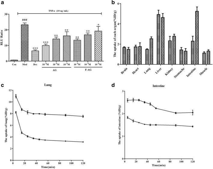
 ) Control (
 ) Model. (c)The lung uptake in the control and model group. (
 ) Control (
 ) Model. (d) The intestine uptake in the control and model groups. (
 ) Control (
 ) Model.
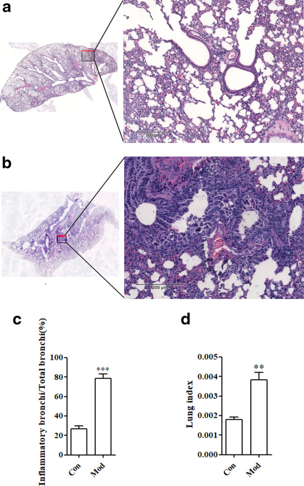

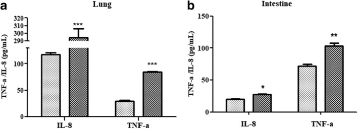
 ) Con (
 ) Mod. (b) Detection of inflammatory factors in the intestine. *
P < 0.05 versus control group; **
P < 0.01 versus control group; ***
P < 0.005 versus control group.
) Mod. (b) Detection of inflammatory factors in the intestine. *
P < 0.05 versus control group; **
P < 0.01 versus control group; ***
P < 0.005 versus control group.Similar articles
-
Andrographolide ameliorates OVA-induced lung injury in mice by suppressing ROS-mediated NF-κB signaling and NLRP3 inflammasome activation.Oncotarget. 2016 Dec 6;7(49):80262-80274. doi: 10.18632/oncotarget.12918. Oncotarget. 2016. PMID: 27793052 Free PMC article.
-
Andrographolide acts as an anti-inflammatory agent in LPS-stimulated RAW264.7 macrophages by inhibiting STAT3-mediated suppression of the NF-κB pathway.J Ethnopharmacol. 2011 Jun 1;135(3):678-84. doi: 10.1016/j.jep.2011.03.068. Epub 2011 Apr 8. J Ethnopharmacol. 2011. PMID: 21497192
-
From irreversible to reversible covalent inhibitors: Harnessing the andrographolide scaffold for anti-inflammatory action.Eur J Med Chem. 2020 Oct 15;204:112481. doi: 10.1016/j.ejmech.2020.112481. Epub 2020 Jul 12. Eur J Med Chem. 2020. PMID: 32712435
-
Andrographolide and its Derivatives in Cardiovascular Disease: A Comprehensive Review.Planta Med. 2025 Apr;91(5):259-270. doi: 10.1055/a-2542-0756. Epub 2025 Mar 7. Planta Med. 2025. PMID: 40054492 Review.
-
Is there a future for andrographolide to be an anti-inflammatory drug? Deciphering its major mechanisms of action.Biochem Pharmacol. 2017 Sep 1;139:71-81. doi: 10.1016/j.bcp.2017.03.024. Epub 2017 Apr 2. Biochem Pharmacol. 2017. PMID: 28377280 Review.
Cited by
-
Exploring the Anticancer Properties of 1,2,3-Triazole-Substituted Andrographolide Derivatives.Pharmaceuticals (Basel). 2025 May 19;18(5):750. doi: 10.3390/ph18050750. Pharmaceuticals (Basel). 2025. PMID: 40430567 Free PMC article.
References
-
- Ye X. Dong MH. A review on different English versions of an ancient classic of Chinese medicine: Huang Di Nei Jing. J Integr Med 2017; 15: 11–8. - PubMed
-
- Phelan AL, Katz R, Gostin LO. The novel coronavirus originating in Wuhan, China: challenges for Global Health governance. JAMA 2020; 323: 709. - PubMed
Publication types
MeSH terms
Substances
Grants and funding
LinkOut - more resources
Full Text Sources
