Review
. 2021 Jan;1868(1):118882.
doi: 10.1016/j.bbamcr.2020.118882. Epub 2020 Oct 2.

Zinc homeostasis and signaling in the roundworm C. elegans

Affiliations
Review

Zinc homeostasis and signaling in the roundworm C. elegans

Brian J Earley et al. Biochim Biophys Acta Mol Cell Res. 2021 Jan.

Abstract

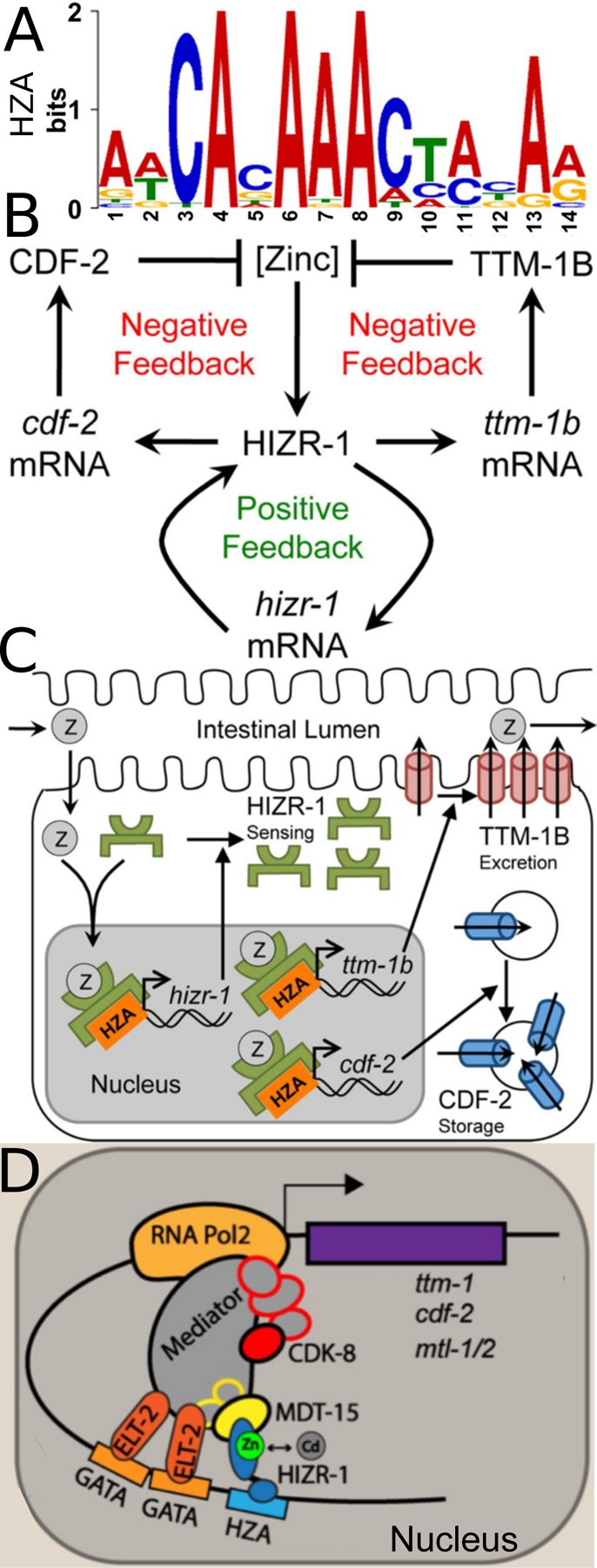
C. elegans is a powerful model for studies of zinc biology. Here we review recent discoveries and emphasize the advantages of this model organism. Methods for manipulating and measuring zinc levels have been developed in or adapted to the worm. The C. elegans genome encodes highly conserved zinc transporters, and their expression and function are beginning to be characterized. Homeostatic mechanisms have evolved to respond to high and low zinc conditions. The pathway for high zinc homeostasis has been recently elucidated based on the discovery of the master regulator of high zinc homeostasis, HIZR-1. A parallel pathway for low zinc homeostasis is beginning to emerge based on the discovery of the Low Zinc Activation promoter element. Zinc has been established to play a role in two cell fate determination events, and accumulating evidence suggests zinc may function as a second messenger signaling molecule during vulval cell development and sperm activation.

Keywords: HIZR-1; LZA; Lysosome-related organelles; Sperm activation; Vulval development; Zinc transporters.


Figures

Figure 1:
Figure 1:. The body plan of C. elegans.
(A) Brightfield images of an adult C. elegans hermaphrodite and a recently laid egg. Scale bar = 0.1 millimeters. (B) Diagram highlighting major anatomical features relevant to zinc biology. The digestive tract begins at the mouth where food enters the pharynx (white). Bacterial food is ground in the pharynx and then enters the intestinal lumen (central line). Nutrients are absorbed from the lumen into intestinal cells (blue), and molecules can be excreted from intestinal cells into the lumen. Defecation occurs at the rectum. The germ line is composed of two gonad arms (purple). From the distal tips of the gonad arms, germ cells travel toward the vulva. During the fourth larval stage of development, hermaphrodite germ cells differentiate into spermatocytes. These spermatocytes are stored in the spermathecae in each gonad arm. Following the fourth larval stage, hermaphrodite germ cells differentiate into oocytes. As each oocyte passes through the spermatheca, the oocyte is fertilized by a spermatocyte and becomes a fertilized embryo. Embryos enter the uterus (fuchsia), where embryos develop a hardened shell and cell divisions progress rapidly. Developing embryos are laid into the environment, at which point they are termed eggs. Within 3 days of hatching, eggs develop into reproductively mature adults.
Figure 2:
Figure 2:. Methods to manipulate dietary zinc and quantify and visualize zinc in C. elegans.
(A) Wild-type worms were cultured in CeMM with different concentrations of added zinc, and maturation was scored by visual inspection. Zinc is necessary for maturation, and high levels of zinc cause toxicity. Adapted from Davis et al., (2009) [12]. (B) Wild type and hizr-1 loss-of-function mutant worms were synchronized as embryos, cultured with the indicated concentrations of supplemental zinc on NAMM for 3 days, and the length of individuals was measured. High levels of zinc inhibit growth, and this is a sensitive method for identifying phenotypic differences in mutants. Adapted from Warnhoff et al., (2017) [33] (C) Embryos were dissected from untreated worms into medium containing 0 (control) or 10 μM TPEN; images are frames from time lapse movies of worms expressing GFP::tubulin and GFP::histone to visualize spindle dynamics. TPEN produces zinc deficient conditions and causes significant delays and abnormalities in cell division. Red arrowheads indicate pronuclei of prophase. Scale bar = 10 μm. Adapted from Mendoza et al., (2017) [15]. (D) Zinc content of mixed-stage wild-type animals. Worms were cultured in CeMM with a range of added zinc, shown on a logarithmic scale. The zinc content was determined by ICP-MS (ppm, closed green circles), or radiolabeled Zn-65 (average ng zinc/µg protein, open green circles). Animals cultured in CeMM with increasing concentrations of zinc display an increase in zinc content. Adapted from Davis et al., (2009) [12]. (E) Fluorescence images of live wild-type hermaphrodites cultured with FluoZin-3 and the indicated levels of supplemental zinc and TPEN. Panels display the anterior half of the intestine of a single animal with pharynx to the left and tail to the right. Scale bar = 50 µm. The FluoZin-3 signal increases with zinc supplementation and decreases with addition of TPEN. Adapted from Roh et al., (2012) [14]. (F) Wild-type animals cultured for 48 hours on NGM with 0 or 500 µM supplemental zinc and assayed for zinc accumulation with spatial resolution by X-ray fluorescence imaging (XFI) at the Stanford Synchrotron Radiation Laboratory. The false color scale indicates the levels of zinc at different positions in the animal. Scale bar = 100 µm. Adapted from Essig et al., (2016) [23].
Figure 3:
Figure 3:. C. elegans CDF and ZIP transporters display orthology with zinc transporters from other species including Homo sapiens.
Phylogenetic trees of CDF (A) and ZIP (B) family members identified by PSI-BLAST from C. elegans (red), Homo sapiens (blue), Arabidopsis thaliana (green), and Saccharomyces cerevisiae (yellow). All ten human ZnT proteins in panel A and all 14 human ZIP proteins in panel B cluster with highly related C. elegans proteins. Transporters discussed in this review are indicated by arrows. Adapted from Roh et al., 2013 [20] (A) and Dietrich et al., (2017) [13] (B).
Figure 4:
Figure 4:. High zinc homeostasis in C. elegans.
(A) A position weight matrix of the High Zinc Activation (HZA) promoter element based on 29 sequences. The height of the nucleotides at each position represents the frequency scaled in bits. Adapted from Roh et al., (2015) [36]. (B) A Genetic model of high zinc homeostasis in the C. elegans intestine. High levels of zinc promote HIZR-1 activity and transcriptional activation of multiple genes including cdf-2, ttm-1b and hizr-1. Increased levels of cdf-2 and ttm-1b mRNA promote increased levels of CDF-2 and TTM-1B protein, which reduce levels of cytoplasmic zinc in a parallel negative feedback circuit. Increased levels of hizr-1 mRNA promote increased levels of HIZR-1 protein, creating a positive feedback circuit that enhances the negative feedback system. Adapted from Warnhoff et al., (2017) [33]. (C) A molecular model. Dietary zinc (Z) enters intestinal cells, binds the ligand-binding domain of HIZR-1, and promotes nuclear accumulation, HZA enhancer binding, and transcriptional activation. The nuclear accumulation of HIZR-1 could result from increased HIZR-1 protein levels due to autoregulation and/ or translocation of HIZR-1 from the cytoplasm to the nucleus. Increased levels of CDF-2 and TTM-1B promote zinc detoxification by sequestration in lysosome-related organelles [14] and excretion into the intestinal lumen [20], respectively. Increased levels of HIZR-1 promote homeostasis by a positive feedback circuit. Adapted from Warnhoff et al., (2017) [33]. (D) A model of the transcriptional activation complex assembled on the cdr-1 promoter in response to high zinc or cadmium. In intestinal cells, MDT-15 cooperates with HIZR-1 bound to the HZA element to induce the metal-sequestering metallothioneins (mtl-1 and −2) and the transporter cdf-2. ELT-2 binds GATA promoter elements to promote intestinal expression of genes and may also contact Mediator to regulate cadmium or zinc responsive transcription. Adapted from Shomer et al., (2019) [37].
Figure 5:
Figure 5:. High Zinc Induces the Formation of Asymmetric Bilobed Gut Granules
(A) Fluorescence images of live transgenic animals expressing CDF-2::GFP cultured with LysoTracker and the indicated levels of supplemental zinc. The differential interference contrast (DIC) images show the intestinal lumen (white triangle) and adjacent intestinal cells with pharynx to the left and tail to the right. Boxed regions are magnified in the right panels. With 200 µM supplemental zinc, many gut granules appear to be bilobed and asymmetric; one side is positive for CDF-2::GFP and LysoTracker (arrowhead), whereas the other side is positive for CDF-2::GFP and negative for LysoTracker (arrow). Scale bars: 10 µm and 2 µm (boxed regions). Adapted from Roh et al., (2012) [14]. (B) A genetic pathway for the formation of bilobed gut granules. The activity of granule biogenesis genes pgp-2 and glo-3 are necessary for the formation of bilobed granules. The zinc transporter cdf-2 is necessary for loading zinc into granules (indicated by dark green). Adapted from Roh et al., (2012) [14].
Figure 6:
Figure 6:. Low zinc homeostasis in C. elegans
(A) A position weight matrix of the LZA element based on 12 sequences. The height of each nucleotide represents the frequency scaled in bits. Adapted from Dietrich et al., (2017) [13]. (B,C) Diagram of the zipt-2.3 promoter containing LZA1 (blue) and LZA2 (red) fused to GFP (green). Images show transgenic animals expressing this construct at the young adult stage cultured on the indicated concentration of TPEN. Bright field images (insets) show worm morphology, and fluorescent images show GFP fluorescence. GFP signals were captured with identical settings and exposure times. zipt-2.3 transcription is activated in response to zinc deficient conditions. Scale bars = 100 µm in brightfield (insets) and fluorescence images. Adapted from Dietrich et al., (2017) [13]. (D) Diagram of an intestinal cell flanked by the intestinal lumen (above) and the pseudocoelom (below). Two dietary zinc-conditions are illustrated: deficiency (left) and excess (right). In the presence of low dietary zinc (black circles), an undefined sensing mechanism causes activation of LZA element containing promoters. ZIP proteins (blue) might localize to the plasma membrane or lysosome related organelles (LRO, green), but the subcellular localization has not been established. In the presence of high dietary zinc, the HIZR-1 nuclear receptor causes activation of HZA element containing promoters. CDF-2 protein (red) localizes to the LRO where it functions to store zinc, and the TTM-1B protein (red) localizes to the apical plasma membrane where it functions to excrete zinc into the intestinal lumen. Adapted from Dietrich et al., (2017) [13]
Figure 7:
Figure 7:. ZIPT-7.1 mediates sperm activation
Model of the biochemical function of ZIPT-7.1 at three times during sperm activation: (1) Primed spermatid: prior to activation, spermatids are primed to respond; ZIPT-7.1 (dark blue) is inactive and localized to the membrane of a vesicle that has a high internal concentration of stored zinc (orange circles) that is presumably the membranous organelle (light blue). The cytoplasm has a low concentration of zinc, and the equilibrium of zinc-regulated proteins (purple) is shifted towards unbound. (2) Release: a sperm-activating signal results in activation of ZIPT-7.1 (orange arrow), zinc begins to flow into the cytoplasm, and zinc-regulated proteins begin binding zinc. (3) Activated spermatozoon: the cytoplasmic concentration of zinc is now high, and zinc-regulated proteins are bound to zinc. The pseudopod extension begins (lower), and the membranous organelles fuse with the plasma membrane. Adapted from Zhao et al., (2018) [21].

Similar articles

Cited by

References

    1. Brenner S, The genetics of Caenorhabditis elegans, Genetics, 77 (1974) 71–94. - PMC - PubMed
    1. Sulston JE, Schierenberg E, White JG, Thomson JN, The embryonic cell lineage of the nematode Caenorhabditis elegans, Dev Biol, 100 (1983) 64–119. - PubMed
    1. Sulston JE, Horvitz HR, Post-embryonic cell lineages of the nematode, Caenorhabditis elegans, Dev Biol, 56 (1977) 110–156. - PubMed
    1. Kimble J, Hirsh D, The postembryonic cell lineages of the hermaphrodite and male gonads in Caenorhabditis elegans, Dev Biol, 70 (1979) 396–417. - PubMed
    1. McGhee JD, The C. elegans intestine, WormBook, (2007) 1–36. - PMC - PubMed

Publication types

MeSH terms

LinkOut - more resources