Rescue endovascular treatment for rapid regrowth of aneurysm remnant on middle cerebral artery trunk after unsuccessful surgical clipping in patients with a ruptured cerebral aneurysm: A report of two cases
- PMID: 33017879
- PMCID: PMC8256017
- DOI: 10.7461/jcen.2020.E2020.08.004
Rescue endovascular treatment for rapid regrowth of aneurysm remnant on middle cerebral artery trunk after unsuccessful surgical clipping in patients with a ruptured cerebral aneurysm: A report of two cases
Abstract
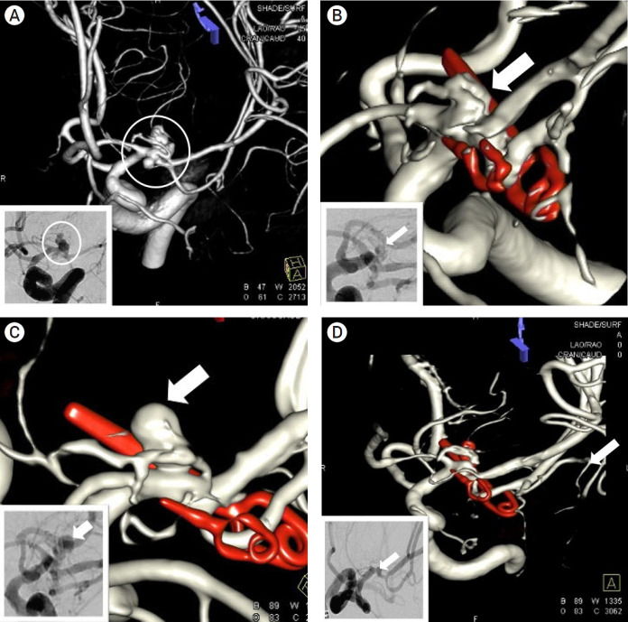
We report two rare cases treated with coiling after rapid regrowth (within a month) of an aneurysm remnant on the middle cerebral artery (MCA) trunk after incomplete surgical clipping. The first case, a 47-year-old man with subarachonoid hemorrhage (SAH) (Hunt-Hess grade II, Fisher grade III) underwent clipping of a ruptured saccular aneurysm with a wide neck on the right early frontal branch arising from the MCA trunk. Incomplete clipping with a 1 mm sized remnant neck was performed to avoid sacrificing the lenticulostriate artery. In a follow-up cerebral angiogram on postoperative day 30, a rapid regrowth of the aneurysm remnant was observed, and on that day, complete obliteration was obtained by rescue endovascular treatment. The second case, a 48-year-old healthy woman with SAH (Hunt-Hess grade II, Fisher grade III) underwent clipping of an anteroposteriorly projecting bilobulated aneurysm on the left M1. Incomplete clipping with a minimal remnant neck was performed. In follow-up digital subtraction angiogram on postoperative day 30, a rapid regrowth of an aneurysm remnant involving only a part of the initial aneurysm near the neck was observed, and on that day, complete obliteration was obtained by rescue coiling. These patients were both discharged without any neurological deficits.
Keywords: Incomplete surgical clipping; Middle cerebral artery trunk; Regrowth of aneurysm remnant; Rescue endovascular treatment.
Figures

References
-
- Burkhardt JK, Chua MHJ, Weiss M, Do AS-MS, Winkler EA, Lawton MT. Risk of aneurysm residual regrowth, recurrence, and de novo aneurysm formation after microsurgical clip occlusion based on follow-up with catheter angiography. World Neurosurg. 2017 Oct;106:74–84. - PubMed
-
- Cekirge HS, Islak C, Firat MM, Kocer N, Saatci I. Endovascular coil embolization of residual or recurrent aneurysms after surgical clipping. Acta Radiol. 2000 Mar;41(2):111–5. - PubMed
-
- Dashti R, Rinne J, Hernesniemi J, Niemelä M, Kivipelto L, Lehecka M, et al. Microneurosurgical management of proximal middle cerebral artery aneurysms. Surg Neurol. 2007 Jan;67(1):6–14. - PubMed
-
- Della Pepa GM, Bianchi F, Scerrati A, Albanese A, Cotroneo E, Delitala A, et al. Secondary coiling after incomplete surgical clipping of cerebral aneurysms: a rescue strategy or a treatment option for complex cases? Institutional series and systematic review. Neurosurg Rev. 2019 Jun;42(2):337–50. - PubMed
-
- El Beltagy M, Muroi C, Roth P, Fandino J, Imhof H-G, Yonekawa Y. Recurrent intracranial aneurysms after successful neck clipping. World Neurosurg. 2010 Oct-Nov;74(4-5):472–7. - PubMed