Past, Present, and Future in the Study of Neural Control of the Lower Urinary Tract
- PMID: 33017890
- PMCID: PMC7538290
- DOI: 10.5213/inj.2040318.159
Past, Present, and Future in the Study of Neural Control of the Lower Urinary Tract
Abstract
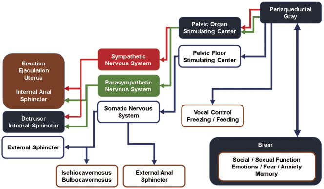
The neurological coordination of the lower urinary tract can be analyzed from the perspective of motor neurons or sensory neurons. First, sensory nerves with receptors in the bladder and urethra transmits stimuli to the cerebral cortex through the periaqueductal gray (PAG) of the midbrain. Upon the recognition of stimuli, the cerebrum carries out decision-making in response. Motor neurons are divided into upper motor neurons (UMNs) and lower motor neurons (LMNs) and UMNs coordinate storage and urination in the brainstem for synergic voiding. In contrast, LMNs, which originate in the spinal cord, cause muscles to contract. These neurons are present in the sacrum, and in particular, a specific neuron group called Onuf's nucleus is responsible for the contraction of the external urethral sphincter and maintains continence in states of rising vesical pressure through voluntary contraction of the sphincter. Parasympathetic neurons originating from S2-S4 are responsible for the contraction of bladder muscles, while sympathetic neurons are responsible for contraction of the urethral smooth muscle, including the bladder neck, during the guarding reflex. UMNs are controlled in the pons where various motor stimuli to the LMNs are directed along with control to various other pelvic organs, and in the PAG, where complex signals from the brain are received and integrated. Future understanding of the complex mechanisms of micturition requires integrative knowledge from various fields encompassing these distinct disciplines.
Keywords: Nervous system; Periaqueductal gray; Pons; Urination.
Conflict of interest statement
No potential conflict of interest relevant to this article was reported.
Figures
Similar articles
-
Sophisticated regulation of micturition: review of basic neurourology.J Exerc Rehabil. 2021 Oct 26;17(5):295-307. doi: 10.12965/jer.2142594.297. eCollection 2021 Oct. J Exerc Rehabil. 2021. PMID: 34805017 Free PMC article. Review.
-
[Lower urinary tract function and its disorders].Cesk Fysiol. 2000 Aug;49(3):134-44. Cesk Fysiol. 2000. PMID: 11039243 Review. Czech.
-
Neuroanatomy, Pontine Micturition Center.2023 Sep 4. In: StatPearls [Internet]. Treasure Island (FL): StatPearls Publishing; 2025 Jan–. 2023 Sep 4. In: StatPearls [Internet]. Treasure Island (FL): StatPearls Publishing; 2025 Jan–. PMID: 32491351 Free Books & Documents.
-
Sensory feedback from the urethra evokes state-dependent lower urinary tract reflexes in rat.J Physiol. 2017 Aug 15;595(16):5687-5698. doi: 10.1113/JP274191. Epub 2017 Jul 7. J Physiol. 2017. PMID: 28612936 Free PMC article.
-
Applied anatomy and physiology of the feline lower urinary tract.Vet Clin North Am Small Anim Pract. 1996 Mar;26(2):181-96. Vet Clin North Am Small Anim Pract. 1996. PMID: 8711856 Review.
Cited by
-
Novel botulinum neurotoxin-A tibial nerve perineural injection to alleviate overactive bladder symptoms in male rats.Anim Cells Syst (Seoul). 2022 Oct 20;26(6):283-290. doi: 10.1080/19768354.2022.2136239. eCollection 2022. Anim Cells Syst (Seoul). 2022. PMID: 36605585 Free PMC article.
-
Research and progress on the mechanism of lower urinary tract neuromodulation: a literature review.PeerJ. 2024 Aug 12;12:e17870. doi: 10.7717/peerj.17870. eCollection 2024. PeerJ. 2024. PMID: 39148679 Free PMC article. Review.
-
Sophisticated regulation of micturition: review of basic neurourology.J Exerc Rehabil. 2021 Oct 26;17(5):295-307. doi: 10.12965/jer.2142594.297. eCollection 2021 Oct. J Exerc Rehabil. 2021. PMID: 34805017 Free PMC article. Review.
-
Bladder Responses to Thoracolumbar Epidural Stimulation in Female Urethane-Anesthetized Rats with Graded Contusion Spinal Cord Injuries.J Neurotrauma. 2025 Feb;42(3-4):229-241. doi: 10.1089/neu.2024.0209. Epub 2024 Sep 26. J Neurotrauma. 2025. PMID: 39264865
-
Diffusion Tensor Imaging: The High-Resolution Image of Functionality in the Central Nervous System.Int Neurourol J. 2022 Sep;26(3):171-172. doi: 10.5213/inj.2222edi03. Epub 2022 Sep 30. Int Neurourol J. 2022. PMID: 36203249 Free PMC article. No abstract available.
References
-
- Vicary T, Furnivall JF, Furnivall P. The anatomie of the bodie of man. London: Early English text society; 1888.
-
- Tortora GJ, Derrickson BH. Principles of anatomy and physiology. 15th ed. Hoboken (NJ): John Wiley & Sons; 2018.
-
- Lim SH, Wang TJ, Tseng GF, Lee YF, Huang YS, Chen JR, et al. The distribution of muscles fibers and their types in the female rat urethra: cytoarchitecture and three-dimensional reconstruction. Anat Rec (Hoboken) 2013;296:1640–9. - PubMed
LinkOut - more resources
Full Text Sources
Medical
Miscellaneous