

Simulating bidirectional peripheral neural interfaces in EIDORS
- PMID: 33018621
- PMCID: PMC9173844
- DOI: 10.1109/EMBC44109.2020.9175921
Simulating bidirectional peripheral neural interfaces in EIDORS
Abstract
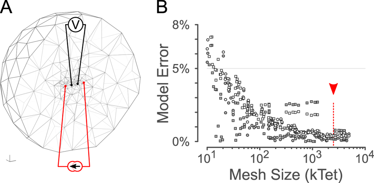
Bioelectronic neural interfaces that deliver adaptive therapeutic stimulation in an intelligent manner must be able to sense and stimulate activity within the same nerve. Existing minimally-invasive peripheral neural interfaces can provide a read-out of the aggregate level of activity via electrical recordings of nerve activity, but these recordings are limited in terms of their specificity. Computational simulations can provide fine-grained insight into the contributions of different neural populations to the extracellular recording, but integration of the signals from individual nerve fibers requires knowledge of spread of current in the complex (heterogenous, anisotropic) extracellular space. We have developed a model which uses the open-source EIDORS package for extracellular stimulation and recording in the pelvic nerve. The pelvic nerve is the primary source of autonomic innervation to the pelvic organs, and a prime target for electrical stimulation to treat a variety of voiding disorders. We simulated recordings of spontaneous and electrically-evoked activity using biophysical models for myelinated and unmyelinated axons. As expected, stimulus thresholds depended strongly on both fibre type and electrode-fibre distance. In conclusion, EIDORS can be used to accurately simulate extracellular recording in complex, heterogenous neural geometries.
Figures


References
-
- Bonaz B, Sinniger V, and Pellissier S, “Vagus nerve stimulation: a new promising therapeutic tool in inflammatory bowel disease,” Journal of Internal Medicine, vol. 282, no. 1, pp. 46–63, 2017. - PubMed
-
- Fall M, “Electrical pelvic floor stimulation for the control of detrusor instability,” Neurourology and Urodynamics, vol. 4, no. 4, pp. 329–335, 1985.
-
- Middleton JW and Keast JR, “Artificial autonomic reflexes: using functional electrical stimulation to mimic bladder reflexes after injury or disease,” Autonomic Neuroscience, vol. 113, no. 1–2, pp. 3–15, 2004. - PubMed
-
- Osborne PB, “Stimulating bioelectronic medicine discovery for urological disorders,” 2017. - PubMed
-
- Durand DM, Grill WM, and Kirsch R, “Electrical stimulation of the neuromuscular system,” in Neural Engineering. Springer, 2005, pp. 157–191.
MeSH terms
Grants and funding
LinkOut - more resources
Full Text Sources
Research Materials