. 2020 Oct 1;8(4):565.
doi: 10.3390/vaccines8040565.

Intradermal Delivery of Dendritic Cell-Targeting Chimeric mAbs Genetically Fused to Type 2 Dengue Virus Nonstructural Protein 1

Affiliations

Intradermal Delivery of Dendritic Cell-Targeting Chimeric mAbs Genetically Fused to Type 2 Dengue Virus Nonstructural Protein 1

Lennon Ramos Pereira et al. Vaccines (Basel). .

Abstract

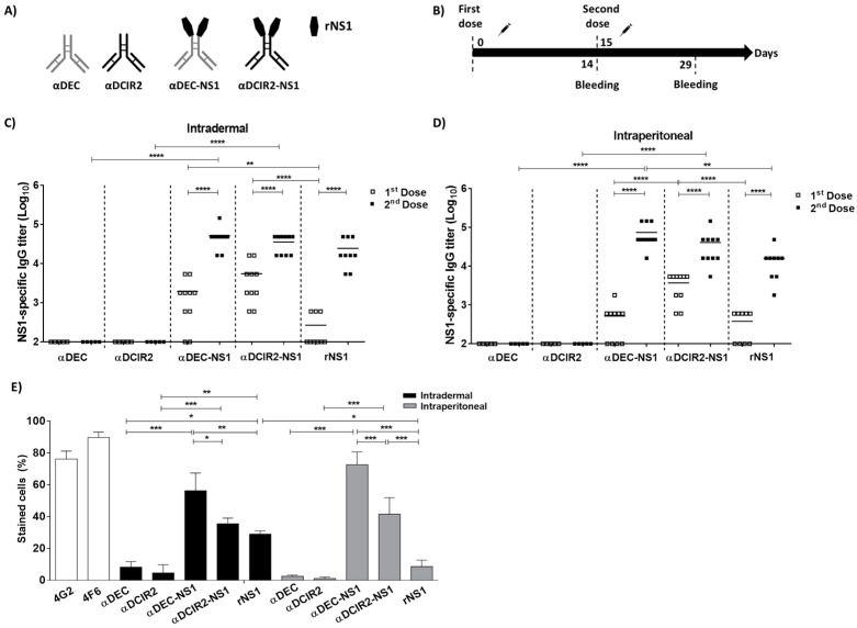
Targeting dendritic cells (DCs) by means of monoclonal antibodies (mAbs) capable of binding their surface receptors (DEC205 and DCIR2) has previously been shown to enhance the immunogenicity of genetically fused antigens. This approach has been repeatedly demonstrated to enhance the induced immune responses to passenger antigens and thus represents a promising therapeutic and/or prophylactic strategy against different infectious diseases. Additionally, under experimental conditions, chimeric αDEC205 or αDCIR2 mAbs are usually administered via an intraperitoneal (i.p.) route, which is not reproducible in clinical settings. In this study, we characterized the delivery of chimeric αDEC205 or αDCIR2 mAbs via an intradermal (i.d.) route, compared the elicited humoral immune responses, and evaluated the safety of this potential immunization strategy under preclinical conditions. As a model antigen, we used type 2 dengue virus (DENV2) nonstructural protein 1 (NS1). The results show that the administration of chimeric DC-targeting mAbs via the i.d. route induced humoral immune responses to the passenger antigen equivalent or superior to those elicited by i.p. immunization with no toxic effects to the animals. Collectively, these results clearly indicate that i.d. administration of DC-targeting chimeric mAbs presents promising approaches for the development of subunit vaccines, particularly against DENV and other flaviviruses.

Keywords: DCIR2; DEC205; Dengue virus; NS1 protein; dendritic cell; intradermal.


Conflict of interest statement

The authors declare no conflict of interest.

Figures

Figure 1
Figure 1
Nonstructural protein 1 (NS1)-specific serum IgG responses in the mice subjected to the tested vaccine regimens. (A) Schematic representation of chimeric monoclonal antibodies (mAbs) and recombinant form of type 2 dengue virus NS1 protein (rNS1). (B) Schematic representation of the tested vaccine regimen. BALB/c mice were immunized via the intradermal (i.d.) or intraperitoneal (i.p.) route with two doses of the vaccine formulations. Serum samples were collected on the indicated days. (C and D) NS1-specific serum IgG titers measured by ELISA following i.d. (C) or i.p. (D) administration. Values represent the individual results (Square symbols) and means (black lines) of the determined IgG titers in log10 scale. (E) Binding of serum antibodies to native NS1 expressed on the DENV2-infected Vero cells. Diluted serum samples collected 14 days after the last dose were incubated with DENV2-infected cells and subsequently analyzed by flow cytometry. The 4G2 and 4F6 mAbs (specific to E and NS1 proteins, respectively) were used as controls. Statistical significance was determined by one- or two-way ANOVA with Bonferroni’s post hoc test (* p < 0.05, ** p < 0.01, *** p < 0.001, **** p < 0.0001).
Figure 2
Figure 2
NS1-specific serum IgG antibody responses in the vaccinated mice. Serum antibodies collected from mice immunized via the i.d. (A) or i.p. (B) routes were evaluated by ELISA using intact (black bars) or heat-denatured (white bars) DENV2 rNS1 protein. The intact/denatured ratios of the IgG titers of each immunization group are indicated on the top of the figure. (C,D) IgG subclass response in the vaccinated mice. NS1-specific serum IgG1, IgG2a, IgG2b, and IgG3 responses were detected in the mice immunized with αDEC-NS1, αDCIR2-NS1, or rNS1 via i.d. (A) or i.p. (B) routes. Serum samples were collected 14 days after the second vaccine dose. Values are expressed as the means ± SD of IgG titers (log10) (n = 5/group). The IgG1/IgG2a ratios of each immunization group are indicated at the top of the figures. (E) Antigen avidity for the NS1-specific antibodies raised in vaccinated mice. Values are expressed as the mean ± SD of the concentration of ammonium thiocyanate required to dissociate 50% of bound anti-NS1 antibodies. Serum samples were collected 14 days after the second immunization dose. Significance was determined by one-way ANOVA with Bonferroni’s post hoc test (* p < 0.05, ** p < 0.01, *** p < 0.001, **** p < 0.0001).
Figure 3
Figure 3
Longevity and boost antibody responses in mice immunized via i.d. and i.p. routes. (A) Schematic representation of a prolonged vaccination schedule. (B,C) NS1-specific IgG titers ± SEM were measured in the pooled serum samples (5 animals/group) collected up to 176 days after the first dose following immunization via the i.d. (B) or i.p. (C) routes. On day 161, mice were boosted with 1 µg of rNS1 using the same administration route. Significance was determined by two-way ANOVA with Bonferroni’s post hoc test (* p < 0.05 comparing rNS1 and αDEC-NS1 groups; # p < 0.05 comparing rNS1 and αDCIR2-NS1 groups; & p < 0.05 comparing αDEC-NS1 and αDCIR2-NS1 groups (see Figure S3 for comparisons between the i.d. and i.p. routes).
Figure 4
Figure 4
Safety parameters of anti-NS1 immune responses elicited in the mice vaccinated with αDEC-NS1, αDCIR2-NS1, or rNS1 via different immunization routes. (A) Binding of anti-NS1 antibodies to platelets in serum samples collected from the mice immunized with αDEC-NS1, αDCIR2-NS1, or rNS1 via the i.d. or i.p. route. The concentration of NS1-specific IgG was adjusted to 10 µg/mL. Platelet binding activity was monitored with an anti-mouse IgG antibody conjugated to Alexa Fluor 488. (B) Platelet aggregation inhibition by NS1-specific serum. Plasma enriched with platelets was incubated with anti-NS1 antibodies collected from the vaccinated mice. After incubation, the platelet suspensions were monitored for aggregation following stimulation with 20 μM adenosine diphosphate (ADP). (C,D) Transendothelial Electrical Resistance (TEER) of human umbilical vein endothelial cells (HUVEC) confluent monolayers incubated with diluted serum samples collected from the mice immunized via i.d. (C) or i.p. (D) routes. TNFα was used as a positive control. TEER was measured at 2, 6, and 24 h after treatment and values expressed as ohms.cm2. Data are showed as the mean ± SEM of two distinct experiments. Significance was determined by one- or two-way ANOVA with Bonferroni’s post hoc test (* p < 0.05).

Similar articles

Cited by

References

    1. Tesfaye D.Y., Gudjonsson A., Bogen B., Fossum E. Targeting Conventional Dendritic Cells to Fine-Tune Antibody Responses. Front. Immunol. 2019;10 doi: 10.3389/fimmu.2019.01529. - DOI - PMC - PubMed
    1. Reuter A., Panozza S.E., Macri C., Dumont C., Li J., Liu H., Segura E., Vega-Ramos J., Gupta N., Caminschi I., et al. Criteria for Dendritic Cell Receptor Selection for Efficient Antibody-Targeted Vaccination. J. Immunol. 2015;194:2696–2705. doi: 10.4049/jimmunol.1402535. - DOI - PubMed
    1. Lehmann C., Heger L., Heidkamp G., Baranska A., Lühr J., Hoffmann A., Dudziak D. Direct Delivery of Antigens to Dendritic Cells via Antibodies Specific for Endocytic Receptors as a Promising Strategy for Future Therapies. Vaccines. 2016;4:8. doi: 10.3390/vaccines4020008. - DOI - PMC - PubMed
    1. Jiang W., Swiggard W.J., Heufler C., Peng M., Mirza A., Steinman R.M., Nussenzweig M.C. The receptor DEC-205 expressed by dendritic cells and thymic epithelial cells is involved in antigen processing. Nature. 1995;375:151–155. doi: 10.1038/375151a0. - DOI - PubMed
    1. Mahnke K., Guo M., Lee S., Sepulveda H., Swain S.L., Nussenzweig M., Steinman R.M. The Dendritic Cell Receptor for Endocytosis, Dec-205, Can Recycle and Enhance Antigen Presentation via Major Histocompatibility Complex Class II–Positive Lysosomal Compartments. J. Cell Biol. 2000;151:673–684. doi: 10.1083/jcb.151.3.673. - DOI - PMC - PubMed