Review
. 2020 Oct 1;21(19):7274.
doi: 10.3390/ijms21197274.

Beta Cell Imaging-From Pre-Clinical Validation to First in Man Testing

Affiliations
Review

Beta Cell Imaging-From Pre-Clinical Validation to First in Man Testing

Stephane Demine et al. Int J Mol Sci. .

Abstract

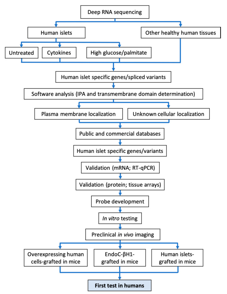
There are presently no reliable ways to quantify human pancreatic beta cell mass (BCM) in vivo, which prevents an accurate understanding of the progressive beta cell loss in diabetes or following islet transplantation. Furthermore, the lack of beta cell imaging hampers the evaluation of the impact of new drugs aiming to prevent beta cell loss or to restore BCM in diabetes. We presently discuss the potential value of BCM determination as a cornerstone for individualized therapies in diabetes, describe the presently available probes for human BCM evaluation, and discuss our approach for the discovery of novel beta cell biomarkers, based on the determination of specific splice variants present in human beta cells. This has already led to the identification of DPP6 and FXYD2ga as two promising targets for human BCM imaging, and is followed by a discussion of potential safety issues, the role for radiochemistry in the improvement of BCM imaging, and concludes with an overview of the different steps from pre-clinical validation to a first-in-man trial for novel tracers.

Keywords: MRI; PET; SPECT; beta cell imaging; pancreas; pre-clinical validation; radiochemistry; type 1 diabetes; type 2 diabetes.


Conflict of interest statement

D.L.E. has patents for the use of FXYD2γα and DPP6 and the tracers targeting them for pancreatic islet cell imaging.

Figures

Figure 1
Figure 1
Step-by-step workflow presently used by our group to identify new beta cell biomarkers and to generate corresponding imaging probes. A schematic overview of the methodology used to mine RNA sequencing data for discovery of novel pancreatic islet biomarkers is shown. Transcripts differentially expressed in pancreatic islets are identified by comparing transcriptomes of human pancreatic islets against 16 different normal human tissues. Deep RNA sequencing analysis (>180 million reads, to allow identification of >80% splice variants) is performed on untreated human islet preparations or after treatment with pro-inflammatory cytokines (IL-1β + IFN-γ or IFN-α) or exposure to metabolic stress (high glucose and/or palmitate) to identify transcripts unaltered by the stressful conditions prevailing in diabetes. A software analysis (Ingenuity pathway analysis (IPA) analysis + transmembrane domain) is conducted on the generated hits to identify membrane-expressed proteins, potentially reachable with a probe. Once the biomarkers identified are validated at the mRNA and protein levels, one or more imaging probes are developed. After complete in vitro validation, the probes are tested for in vivo imaging on humanize mouse models grafted with different amounts of human beta cells.
Figure 2
Figure 2
SPECT imaging of human islets grafted in immunodeficient mice using an anti-DPP6 nanobody. Female NOD-SCID mice were transplanted subcutaneously in the intrascapular area with different numbers of primary human islets (1000 or 3000 islet equivalent (IEQ)) as described [101]. After 4 weeks, the mice were imaged 60 min post-injection by full body CT followed by a focal single-photon emission computed tomography (SPECT) imaging scan using an 99mTc-anti-DPP6 nanobody. The yellow square indicates the field-of-view of the SPECT camera. The white arrows indicate the graft localization. Full analyses and quantification of these pictures are described in [101].
Figure 3
Figure 3
The block diagrams (A) is the most simplistic model achievable and has been used to describe trappable tracers for blood flow determination such as microspheres [192] (Equation (6)), while (B) represents a similar model, but the tracer is not trapped in the C1 compartment, thus allowing efflux back into the blood as with radioactive water studies [178] (Equation (7)). In diagram (C), the model has been used to describe the binding of ligands to receptor systems, where the C1 compartment represents the interstitial space, and C2 is the receptor complex on the tissue of interest, such as for glucose metabolism [193] or P2X7 receptor binding [173] (Equations (8) and (9)). Lastly, (D) has been used to describe binding to the receptor system described in (C), but the C3 compartment represents the non-specific binding (Equations (10)–(12)) [194].

References

    1. IDF Diabetes Atlas. [(accessed on 25 January 2020)]; Available online: http://www.diabetesatlas.org/
    1. Li S., Wang J., Zhang B., Li X., Liu Y. Diabetes Mellitus and Cause-Specific Mortality: A Population-Based Study. Diabetes Metab. J. 2019;43:319–341. doi: 10.4093/dmj.2018.0060. - DOI - PMC - PubMed
    1. Raghavan S., Vassy J.L., Ho Y.L., Song R.J., Gagnon D.R., Cho K., Wilson P.W.F., Phillips L.S. Diabetes Mellitus-Related All-Cause and Cardiovascular Mortality in a National Cohort of Adults. J. Am. Heart Assoc. 2019;8:e011295. doi: 10.1161/JAHA.118.011295. - DOI - PMC - PubMed
    1. Afkarian M., Sachs M.C., Kestenbaum B., Hirsch I.B., Tuttle K.R., Himmelfarb J., de Boer I.H. Kidney disease and increased mortality risk in type 2 diabetes. J. Am. Soc. Nephrol. 2013;24:302–308. doi: 10.1681/ASN.2012070718. - DOI - PMC - PubMed
    1. Hippisley-Cox J., Coupland C. Diabetes treatments and risk of amputation, blindness, severe kidney failure, hyperglycaemia, and hypoglycaemia: Open cohort study in primary care. BMJ. 2016;352:i1450. doi: 10.1136/bmj.i1450. - DOI - PMC - PubMed

MeSH terms