Distal Displacement of Maxillary Sinus Anterior Wall Versus Conventional Sinus Lift with Lateral Access: A 3-Year Retrospective Computerized Tomography Study
- PMID: 33019711
- PMCID: PMC7579181
- DOI: 10.3390/ijerph17197199
Distal Displacement of Maxillary Sinus Anterior Wall Versus Conventional Sinus Lift with Lateral Access: A 3-Year Retrospective Computerized Tomography Study
Abstract
Background: The present study is designed to compare the outcomes of two sinus augmentation procedures: distal displacement of the anterior wall versus standard sinus lifting and grafting with a lateral window approach.
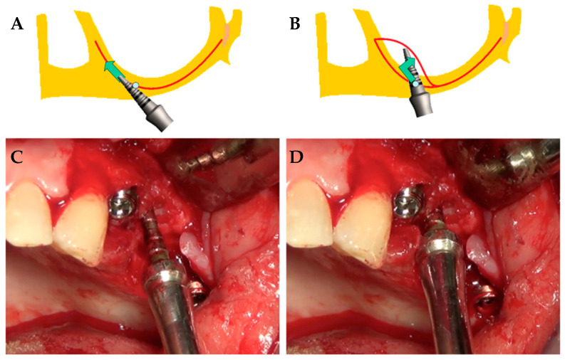
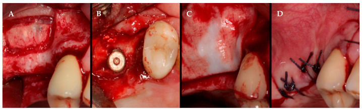
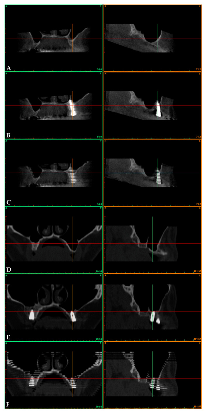
Methods: In the displacement group, a localized surgical fracture of the sinus floor achieved through an electromagnetic device results in the distal displacement of the anterior wall. In the filling group, sinus lifting (with lateral access) and grafting with particulate xenogeneic bone substitute was performed. Bone volume beneath the maxillary sinus was investigated with computerized tomography after baseline and postoperative data superimposition. Clinical and radiological outcomes over three years had been evaluated.
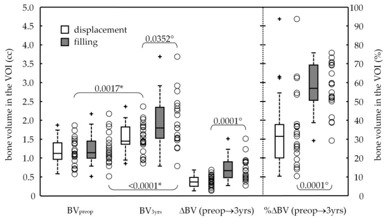
Results: Forty-three dental implants were selected. The two sinus lift procedures significantly increased the bone volume (p-value ≤ 0.0017) in the displacement group from 1.17 ± 0.34 to 1.53 ± 0.39 cc, with a final bone gain of +0.36 ± 0.17 cc, and in the filling group from 1.24 ± 0.41 to 1.94 ± 0.68 cc, with a bone augmentation of +0.71 ± 0.31 cc. No events of dental implant bulging into the maxillary sinus occurred. Two implants failed early on in the filling group, attesting the 3-year survival rate of 92.6% (CI95%: 82.7-100%). Marginal bone loss at the distal aspect was 1.66 ± 0.72 and 1.25 ± 0.78 mm, respectively, for the displacement and filling groups, with a significant difference (p-value = 0.0497).
Conclusion: Results showed a significant and effective bone gain around dental implants at a 3-year survey for both sinus augmented by backward displacement of the anterior wall (+34%) and sinus lifting and grafting with a lateral window approach (+57%).
Keywords: CT imaging; bone augmentation; dental implant; infracture approach; maxillary sinus.
Conflict of interest statement
The authors declare no conflict of interest.
Figures


Similar articles
-
Bone Graft Displacement After Maxillary Sinus Floor Augmentation With or Without Covering Barrier Membrane: A Retrospective Computed Tomographic Image Evaluation.Int J Oral Maxillofac Implants. 2019 May/June;34(3):681–691. doi: 10.11607/jomi.6940. Epub 2018 Dec 5. Int J Oral Maxillofac Implants. 2019. PMID: 30521657
-
Lateral Displacement of Maxillary Sinus Palatal Wall: A 1-Year Retrospective Computerized Tomography Study.Int J Oral Maxillofac Implants. 2022 Sep-Oct;37(5):920-928. doi: 10.11607/jomi.9594. Int J Oral Maxillofac Implants. 2022. PMID: 36170306
-
A 5-year clinical and computerized tomographic implant follow-up in sinus-lifted maxillae and native bone.Clin Oral Implants Res. 2014 Sep;25(9):1056-64. doi: 10.1111/clr.12222. Epub 2013 Jul 4. Clin Oral Implants Res. 2014. PMID: 23822163
-
Lateral trap-door window approach with maxillary sinus membrane lifting for dental implant placement in atrophied edentulous alveolar ridge.J Chin Med Assoc. 2015 Feb;78(2):85-8. doi: 10.1016/j.jcma.2014.05.016. Epub 2014 Oct 3. J Chin Med Assoc. 2015. PMID: 25287252 Review.
-
Molecular, Cellular and Pharmaceutical Aspects of Bone Grafting Materials and Membranes During Maxillary Sinus-lift Procedures. Part 1: A General Overview.Curr Pharm Biotechnol. 2017;18(1):19-32. doi: 10.2174/1389201017666161221155237. Curr Pharm Biotechnol. 2017. PMID: 28003001 Review.
Cited by
-
Biomolecular, Histological, Clinical, and Radiological Analyses of Dental Implant Bone Sites Prepared Using Magnetic Mallet Technology: A Pilot Study in Animals.Materials (Basel). 2021 Nov 17;14(22):6945. doi: 10.3390/ma14226945. Materials (Basel). 2021. PMID: 34832347 Free PMC article.
-
Application Effect of External and Internal Elevation of Maxillary Sinus in Implant Restoration of Posterior Maxilla.Emerg Med Int. 2022 Sep 1;2022:7879633. doi: 10.1155/2022/7879633. eCollection 2022. Emerg Med Int. 2022. Retraction in: Emerg Med Int. 2023 Aug 23;2023:9878673. doi: 10.1155/2023/9878673. PMID: 36090544 Free PMC article. Retracted.
-
Two-Year Follow-Up of 4-mm-Long Implants Used as Distal Support of Full-Arch FDPs Compared to 10-mm Implants Installed after Sinus Floor Elevation. A Randomized Clinical Trial.Int J Environ Res Public Health. 2021 Apr 6;18(7):3846. doi: 10.3390/ijerph18073846. Int J Environ Res Public Health. 2021. PMID: 33917587 Free PMC article. Clinical Trial.
-
Comparison between Magneto-Dynamic, Piezoelectric, and Conventional Surgery for Dental Extractions: A Pilot Study.Dent J (Basel). 2023 Feb 23;11(3):60. doi: 10.3390/dj11030060. Dent J (Basel). 2023. PMID: 36975557 Free PMC article.
-
Effect of Implant Macro-Design and Magnetodynamic Surgical Preparation on Primary Implant Stability: An In Vitro Investigation.Dent J (Basel). 2023 Sep 24;11(10):227. doi: 10.3390/dj11100227. Dent J (Basel). 2023. PMID: 37886912 Free PMC article.
References
-
- Khoury F. Augmentation of the sinus floor with mandibular bone block and simultaneous implantation: A 6-year clinical investigation. Int. J. Oral Maxillofac. Implant. 1999;14:557–564. - PubMed
-
- Boyne P.J., James R.A. Grafting of the maxillary sinus floor with autogenous marrow and bone. J. Oral Surg. 1980;38:613–616. - PubMed
MeSH terms
Substances
LinkOut - more resources
Full Text Sources