. 2020 Nov 15;205(10):2861-2872.
doi: 10.4049/jimmunol.2000810. Epub 2020 Oct 5.

Peptide Binding to HLA-E Molecules in Humans, Nonhuman Primates, and Mice Reveals Unique Binding Peptides but Remarkably Conserved Anchor Residues

Affiliations

Peptide Binding to HLA-E Molecules in Humans, Nonhuman Primates, and Mice Reveals Unique Binding Peptides but Remarkably Conserved Anchor Residues

Paula Ruibal et al. J Immunol. .

Abstract

Ag presentation via the nonclassical MHC class Ib molecule HLA-E, with nearly complete identity between the two alleles expressed in humans, HLA-E*01:01 and HLA-E*01:03, can lead to the activation of unconventional T cells in humans. Despite this virtual genetic monomorphism, differences in peptide repertoires binding to the two allelic variants have been reported. To further dissect and compare peptide binding to HLA-E*01:01 and HLA-E*01:03, we used an UV-mediated peptide exchange binding assay and an HPLC-based competition binding assay. In addition, we investigated binding of these same peptides to Mamu-E, the nonhuman primate homologue of human HLA-E, and to the HLA-E-like molecule Qa-1b in mice. We next exploited the differences and homologies in the peptide binding pockets of these four molecules to identify allele specific as well as common features of peptide binding motifs across species. Our results reveal differences in peptide binding preferences and intensities for each human HLA-E variant compared with Mamu-E and Qa-1b Using extended peptide libraries, we identified and refined the peptide binding motifs for each of the four molecules and found that they share main anchor positions, evidenced by conserved amino acid preferences across the four HLA-E molecules studied. In addition, we also identified differences in peptide binding motifs, which could explain the observed variations in peptide binding preferences and affinities for each of the four HLA-E-like molecules. Our results could help with guiding the selection of candidate pathogen-derived peptides with the capacity to target HLA-E-restricted T cells that could be mobilized in vaccination and immunotherapeutic strategies.


Conflict of interest statement

The authors have no financial conflicts of interest.

Figures
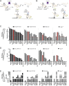
FIGURE 1.
FIGURE 1.
Peptide binding to HLA-E*01:01, HLA-E*01:03, Mamu-E, and Qa-1b quantified with an UV-mediated peptide exchange assay and a competition-based HPLC assay. (A) Schematic representation of the UV-mediated peptide exchange assay to measure peptide binding to HLA-E*01:01 and HLA-E*01:03 by capturing rescued complexes with plates coated with a specific HLA-E Ab. (B) Schematic representation of a modified UV-mediated peptide exchange assay to measure peptide binding to Mamu-E and Qa-1b in which the capture of previously biotinylated complexes is done with streptavidin-coated plates. In both (A and B), rescued complexes are detected with an HRP-coupled β2m Ab. (CE) Previously described MHC-E binding peptides in humans: pCMV (VLAPRTLLL, red bar) and VL9, canonical HLA-E binders; p34-p68, M. tuberculosis–derived peptides previously predicted to bind HLA-E (7); in mice, Qdm-FL9, Qa-1b binders (12); and in NHP, Gag120-Gag69, SIV-derived peptides known to bind Mamu-E (15). The sequences of these peptides are available in Supplemental Table I. Peptides are ranked according to their capacity to bind HLA-E*01:01 to facilitate comparison between alleles. (C) Absorbance represents the number of MHC-E complexes that are rescued in the presence of test peptide, the value being higher for peptides with better binding capacity. Bars show the average and SD of a representative experiment with two biological repeats and two technical replicates. (D) The s/p ratios were calculated by normalizing values to positive control (pCMV) after subtraction of background obtained in the absence of test peptide (no rescue): (value − no rescue)/(pCMV − no rescue). Bars represent calculations from one representative experiment containing two biological repeats and two technical replicates. (E) IC50 were obtained by HPLC size exclusion chromatography using recombinant MHC-E and competition of a fluorescently labeled peptide. Binding was calculated as the concentration (micromolar) of peptide required to reduce fluorescence intensity of the standard peptide by 50% (IC50), and 100 μM was the highest concentration tested. Bars represent one representative experiment.
FIGURE 2.
FIGURE 2.
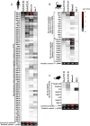
Peptides bind differently to HLA-E*01:01, HLA-E*01:03, Mamu-E, and Qa-1b. Peptide binding was measured using the UV-mediated peptide exchange assay. Heatmaps represent mean values of at least two independent experiments and indicate s/p ratios of the tested peptides for each MHC-E molecule with white being no binding (s/p ratio = 0), black being a binding similar to positive control (s/p ratio = 1), and red being a binding higher than the positive control. Peptides were arranged in a decreasing order of binding to HLA-E*01:01, with the stronger binder at the top and canonical and control binders at the bottom. (A) M. tuberculosis–derived peptides previously predicted to bind HLA-E (7). (B) HIV- and SIV-derived peptides previously described to bind Mamu-E (15, 42, 43). (C) Peptides previously described to bind Qa-1b (13).
FIGURE 3.
FIGURE 3.
Good correlation between UV-mediated peptide exchange and HPLC binding assays. Correlations between the s/p ratio obtained with the UV-mediated peptide exchange binding assay and the IC50 obtained with the HPLC binding assay are plotted for HLA-E*01:03, HLA-E*01:01, Mamu-E, and Qa-1b as indicated. IC50 values for peptides represented as black circles correspond to HIV- and SIV-derived peptides previously described to bind Mamu-E (15, 42, 43) and peptides previously described to bind Qa-1b (13). IC50 values for peptides represented as gray triangles correspond to those previously published in the context of only HLA-E*01:03 (7). Dotted lines represent the thresholds indicating binders and strong binders to each MHC-E molecule, according to the UV-mediated peptide exchange binding assay. The s/p ratios represent mean values from at least two independent experiments with two biological replicates and two technical replicates. IC50 values represent one experiment. Statistical significance was tested by nonparametric Spearman correlation analysis.
FIGURE 4.
FIGURE 4.
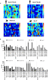
Binding of alanine substitutions reveals the importance of anchor positions for binding HLA-E*01:01, HLA-E*01:03, Mamu-E, and Qa-1b. Peptides were modified by an alanine substitution at each position and tested for binding to HLA-E*01:01, HLA-E*01:03, Mamu-E, and Qa-1b with the UV-mediated peptide exchange binding assay. Each graph represents peptide binding as s/p ratio for a peptide and its alanine substitutions in the context of the four MHC-E molecules. The graphs show the mean and SD of at least two independent experiments with two biological replicates and two technical replicates each.
FIGURE 5.
FIGURE 5.
New insights into amino acid preferences for peptide binding to HLA-E*01:01, HLA-E*01:03, Mamu-E, and Qa-1b. (A) Heatmap representation of CPL binding intensity to HLA-E*01:01, HLA-E*01:03, Mamu-E, and Qa-1b, indicating which amino acids (columns) are preferred at each peptide position (row) for the peptide to bind each MHC-E molecule. Data represent mean values of two independent experiments with two biological replicates and are normalized for each position with values ranging from high binding in red to low binding in blue. Highlighted with black squares are the residues and positions that resulted in preferred binding to each molecule. (B) Amino acid substitutions on pCMV and Mtb44 were tested based on the results above to confirm flexibility or strictly preferred amino acids in anchor positions. Bar graphs show mean and SD from at least two independent experiments. The s/p ratios represent binding affinity of the indicated peptides to HLA-E*01:01, HLA-E*01:03, Mamu-E, and Qa-1b.
FIGURE 6.
FIGURE 6.
Binding motifs to HLA-E*01:01, HLA-E*01:03, Mamu-E, and Qa-1b. New insights into the residue preferences at each peptide position allowing peptide binding to HLA-E*01:01, HLA-E*01:03, Mamu-E, and Qa-1b are indicated. The figure represents a summary of the peptide binding motifs for each MHC-E molecule based on all the data obtained in this study. The canonical sequence of pCMV is indicated with the underlined positions following the motifs. Residues in bold highlight stronger preferences, whereas those in gray are less strongly preferred. Primary anchor positions are highlighted in red, and secondary anchor positions are highlighted in green. Positions with newly defined residue preferences are highlighted in yellow and blue.

Similar articles

Cited by

References

    1. D’Souza M. P., Adams E., Altman J. D., Birnbaum M. E., Boggiano C., Casorati G., Chien Y. H., Conley A., Eckle S. B. G., Früh K., et al. 2019. Casting a wider net: immunosurveillance by nonclassical MHC molecules. PLoS Pathog. 15: e1007567. - PMC - PubMed
    1. Joosten S. A., Ottenhoff T. H. M., Lewinsohn D. M., Hoft D. F., Moody D. B., Seshadri C., Collaboration for Tuberculosis Vaccine Discovery - Donor-Unrestricted T-cells Working Group, Bill and Melinda Gates Foundation 2019. Harnessing donor unrestricted T-cells for new vaccines against tuberculosis. Vaccine 37: 3022–3030. - PMC - PubMed
    1. Geraghty D. E., Stockschleader M., Ishitani A., Hansen J. A. 1992. Polymorphism at the HLA-E locus predates most HLA-A and -B polymorphism. Hum. Immunol. 33: 174–184. - PubMed
    1. Braud V. M., Allan D. S., O’Callaghan C. A., Söderström K., D’Andrea A., Ogg G. S., Lazetic S., Young N. T., Bell J. I., Phillips J. H., et al. 1998. HLA-E binds to natural killer cell receptors CD94/NKG2A, B and C. Nature 391: 795–799. - PubMed
    1. Lee N., Llano M., Carretero M., Ishitani A., Navarro F., López-Botet M., Geraghty D. E. 1998. HLA-E is a major ligand for the natural killer inhibitory receptor CD94/NKG2A. Proc. Natl. Acad. Sci. USA 95: 5199–5204. - PMC - PubMed

Publication types

MeSH terms