. 2020 Dec 11;295(50):17138-17147.
doi: 10.1074/jbc.RA120.014420. Epub 2020 Oct 5.

Microtubule affinity-regulating kinase 4 with an Alzheimer's disease-related mutation promotes tau accumulation and exacerbates neurodegeneration

Affiliations

Microtubule affinity-regulating kinase 4 with an Alzheimer's disease-related mutation promotes tau accumulation and exacerbates neurodegeneration

Toshiya Oba et al. J Biol Chem. .

Abstract

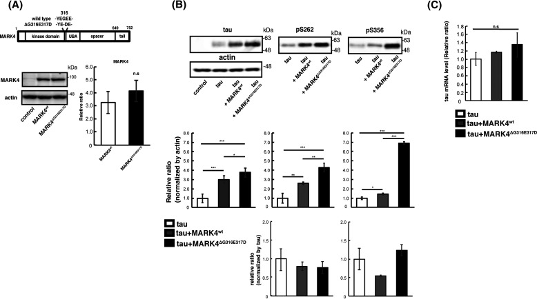
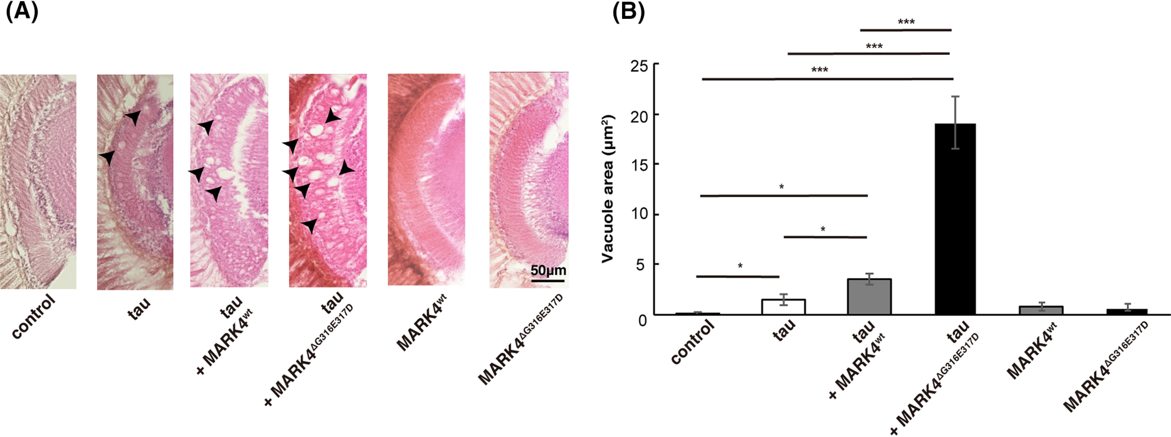
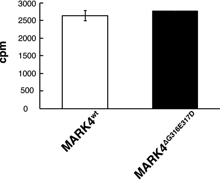
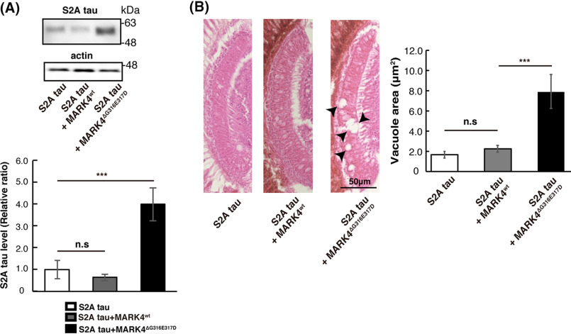
Accumulation of the microtubule-associated protein tau is associated with Alzheimer's disease (AD). In AD brain, tau is abnormally phosphorylated at many sites, and phosphorylation at Ser-262 and Ser-356 plays critical roles in tau accumulation and toxicity. Microtubule affinity-regulating kinase 4 (MARK4) phosphorylates tau at those sites, and a double de novo mutation in the linker region of MARK4, ΔG316E317D, is associated with an elevated risk of AD. However, it remains unclear how this mutation affects phosphorylation, aggregation, and accumulation of tau and tau-induced neurodegeneration. Here, we report that MARK4ΔG316E317D increases the abundance of highly phosphorylated, insoluble tau species and exacerbates neurodegeneration via Ser-262/356-dependent and -independent mechanisms. Using transgenic Drosophila expressing human MARK4 (MARK4wt) or a mutant version of MARK4 (MARK4ΔG316E317D), we found that coexpression of MARK4wt and MARK4ΔG316E317D increased total tau levels and enhanced tau-induced neurodegeneration and that MARK4ΔG316E317D had more potent effects than MARK4wt Interestingly, the in vitro kinase activities of MARK4wt and MARK4ΔG316E317D were similar. When tau phosphorylation at Ser-262 and Ser-356 was blocked by alanine substitutions, MARK4wt did not promote tau accumulation or exacerbate neurodegeneration, whereas coexpression of MARK4ΔG316E317D did. Both MARK4wt and MARK4ΔG316E317D increased the levels of oligomeric forms of tau; however, only MARK4ΔG316E317D further increased the detergent insolubility of tau in vivo Together, these findings suggest that MARK4ΔG316E317D increases tau levels and exacerbates tau toxicity via a novel gain-of-function mechanism and that modification in this region of MARK4 may affect disease pathogenesis.

Keywords: Alzheimer's disease (AD); Drosophila; microtubule affinity–regulating kinase 4 (MARK4); neurodegeneration; phosphorylation; tau protein (tau).


Conflict of interest statement

Conflict of interest—The authors declare that they have no conflicts of interest with the contents of this article.

Figures

Figure 1.
Figure 1.
MARK4ΔG316E317D increases the level of tau to a greater extent than MARK4wt. A, (top) schematic representation of MARK4 and ΔG316E317D mutation. (bottom) MARK4wt and MARK4ΔG316E317D are expressed at similar levels in fly eyes. MARK4 expression was driven by the pan-retinal driver GMR-Gal4. Levels of MARK4wt or MARK4ΔG316E317D in fly head lysates were examined by Western blotting. Actin was used as a loading control. Representative blots (left panels) and quantification (right panels) are shown. B, coexpression of MARK4wt increased the levels of phosphorylated tau and total tau, and coexpression of MARK4ΔG316E317D increased the levels of total tau, pSer-262–tau, and pSer-356–tau in the eyes to a significantly greater extent than MARK4wt. Western blotting was performed with anti-tau antibody T46 (tau), anti-phospho–Ser-262 tau antibody (pSer-262), or anti-phospho–Ser-356 antibody (pSer-356). Representative blots (top) and quantification normalized to actin, or to total tau (bottom panels), are shown. C, coexpression of MARK4wt or MARK4ΔG316E317D does not affect tau mRNA levels. mRNA levels of tau expressed alone (tau), tau coexpressed with MARK4wt (tau+MARK4wt), and tau coexpressed with MARK4ΔG316E317D (tau+MARK4ΔG316E317D) were measured by quantitative PCR. Means ± S.D. (error bars); n = 4. n.s. (not significant), p > 0.05; *p < 0.05; **p < 0.01; ***p < 0.005 (one-way ANOVA and Tukey's post-hoc test).
Figure 2.
Figure 2.
MARK4ΔG316E317D increases tau toxicity to a greater extent than MARK4wt. A, coexpression of MARK4 increased tau-induced neurodegeneration, and MARK4ΔG316E317D exerted a stronger effect than MARK4wt. Shown are lamina of flies carrying GMR-Gal4 driver alone (control), flies expressing tau (tau), flies coexpressing tau and MARK4wt (tau+MARK4wt), or flies coexpressing tau and MARK4ΔG316E317D (tau+MARK4ΔG316E317D). Expression of MARK4wt (MARK4wt) or MARK4ΔG316E317D (MARK4ΔG316E317D) alone did not cause neurodegeneration. Neurodegeneration is observed as vacuoles, indicated by arrows. B, quantification of the vacuole area. Means ± S.E. (error bars). n = 5. n.s. (not significant), p > 0.05; *p < 0.05; **p < 0.01; ***p < 0.005 (one-way ANOVA and Tukey's post-hoc test).
Figure 3.
Figure 3.
MARK4wt or MARK4ΔG316E317D have similar kinase activity in vitro. MARK4wt and MARK4ΔG316E317D expressed in HEK293 cells were immunoprecipitated and subjected to in vitro kinase assays. Incorporation of 32P in recombinant tau protein is expressed as means ± S.D. (error bars). (n = 3, n.s. (not significant), p > 0.05 (Student's t test)).
Figure 4.
Figure 4.
MARK4ΔG316E317D increases tau levels and tau toxicity in a Ser-262/356–independent manner. A, MARK4ΔG316E317D positively regulated tau levels in a Ser-262/356–independent manner. Western blots of fly heads expressing S2A tau alone or coexpressing S2A tau with either MARK4wt (S2A tau+MARK4wt) or MARK4ΔG316E317D (S2A tau+MARK4ΔG316E317D). Means ± S.D. (error bars), n = 4. n.s. (not significant), p > 0.05; ***p < 0.005 (one-way ANOVA and Tukey's post-hoc test). B, coexpression of S2A tau with MARK4ΔG316E317D, but not with MARK4wt, enhanced neurodegeneration. Shown are lamina of flies harboring expressing S2A tau (S2A tau), coexpressing S2A tau and MARK4wt (S2A tau+MARK4wt), or coexpressing S2A tau and MARK4ΔG316E317D (S2A tau+MARK4ΔG316E317D). Means ± S.E. (error bars); n = 5. n.s. (not significant), p > 0.05; ***p < 0.005 (one-way ANOVA and Tukey's post-hoc test).
Figure 5.
Figure 5.
Neither MARK4wt nor MARK4ΔG316E317D inhibit the TOR pathway or increase autophagic activity. A, MARK4ΔG316E317D did not increase the levels of luciferase coexpressed in fly eyes. Western blots were performed on fly heads expressing luciferase alone (luciferase), coexpressing luciferase and MARK4wt (luciferase+MARK4wt), or coexpressing luciferase and MARK4ΔG316E317D (luciferase+MARK4ΔG316E317D). Actin was used as a loading control. Representative blots and quantitation are shown. Means ± S.D. (error bars); n = 4. n.s. (not significant), p > 0.05; *p < 0.05; **p < 0.01; ***p < 0.005 (one-way ANOVA and Tukey's post-hoc test). BD, expression of MARK4wt or MARK4ΔG316E317D did not promote autophagy. Western blots were performed on fly heads expressing control, MARK4wt, or MARK4ΔG316E317D. Blots were performed with anti-pS6K (pS6K) (B), anti-Ref2P (Ref2P) (C), and LC3 antibody (LC3) (D). Representative blots and quantitation are shown. Means ± S.D. (error bars); n = 3. n.s. (not significant), p > 0.05 (one-way ANOVA and Tukey post-hoc test). Actin was used as a loading control.
Figure 6.
Figure 6.
Coexpression of MARK4ΔG316E317D causes accumulation of total tau and tau-phosphorylated at sites other than Ser-262/356 in vivo. A, phosphorylation profile of tau expressed alone or coexpressed with MARK4wt or MARK4ΔG316E317D in vivo. Fly head extracts were separated by Phos-tag SDS-PAGE, followed by Western blotting with anti-tau antibody. Coexpression of MARK4wt increased the abundance of faster-migrating tau. By contrast, coexpression of MARK4ΔG316E317D increased the abundance of slower-migrating tau. B, Western blotting was performed using phospho-specific antibodies for SP/TP sites such as AT8 (pS202/T205), anti-pThr231 (pT231), and PHF1 (pS396/404), as well as pan-tau antibody T46 (tau). Actin was used as a loading control. Representative blots (top) and quantification (bottom panels, normalized either to actin or to total tau) are shown. Means ± S.D. (error bars); n = 4. n.s. (not significant), p > 0.05; *p < 0.05; **p < 0.01; ***p < 0.005 (one-way ANOVA and Tukey's post-hoc test). C, MARK4wt and MARK4ΔG316E317D phosphorylated tau in the same pattern in vitro. MARK4wt and MARK4ΔG316E317D expressed in HEK293 cells were immunoprecipitated and incubated with recombinant tau. Tau proteins were separated by Phos-tag SDS-PAGE, followed by Western blotting with anti-tau antibody T46.
Figure 7.
Figure 7.
MARK4ΔG316E317D promotes misfolding of tau. A, MARK4wt and MARK4ΔG316E317D promoted accumulation of tau oligomers and dimers at similar levels. Western blotting was performed on fly head lysates prepared under nonreducing conditions with oligomer-specific antibody T22 (oligomer) and pan-tau antibody Tau5 (dimer). Representative blots and quantification are shown. Actin was used as a loading control. Means ± S.D. (error bars); n = 3. n.s. (not significant), p > 0.05; *p < 0.05, **p < 0.01 (one-way ANOVA and Tukey's post-hoc test). B and C, tau in extracts from heads expressing tau alone (tau), coexpressing tau and MARK4wt (tau+MARK4wt), or coexpressing tau and MARK4ΔG316E317D (tau+MARK4ΔG316E317D) were detected by Western blotting with pan-tau antibody T46. Head homogenates were extracted first with RIPA buffer containing NP-40 (NP-40), next with 2% SDS (SDS), and finally with 70% formic acid (FA). B, representative Western blots are shown (shorter exposure (upper panel) and longer exposure (lower panel)). C, Western blotting was performed on fly heads extracted with RIPA buffer containing NP-40 (NP-40) and then with 2% SDS (SDS). Representative blots (left) and quantification (right) are shown.

References

    1. Wang, Y., and Mandelkow, E. (2016) Tau in physiology and pathology. Nat. Rev. Neurosci. 17, 5–21 10.1038/nrn.2015.1 - DOI - PubMed
    1. Grundke-Iqbal, I., Iqbal, K., Tung, Y. C., Quinlan, M., Wisniewski, H. M., and Binder, L. I. (1986) Abnormal phosphorylation of the microtubule-associated protein tau (tau) in Alzheimer cytoskeletal pathology. Proc. Natl. Acad. Sci. U. S. A. 83, 4913–4917 10.1073/pnas.83.13.4913 - DOI - PMC - PubMed
    1. Hasegawa, M., Morishima-Kawashima, M., Takio, K., Suzuki, M., Titani, K., and Ihara, Y. (1992) Protein sequence and mass spectrometric analyses of tau in the Alzheimer's disease brain. J. Biol. Chem. 267, 17047–17054 - PubMed
    1. Hanger, D. P., Betts, J. C., Loviny, T. L., Blackstock, W. P., and Anderton, B. H. (1998) New phosphorylation sites identified in hyperphosphorylated tau (paired helical filament-tau) from Alzheimer's disease brain using nanoelectrospray mass spectrometry. J. Neurochem. 71, 2465–2476 10.1046/j.1471-4159.1998.71062465.x - DOI - PubMed
    1. Morishima-Kawashima, M., Hasegawa, M., Takio, K., Suzuki, M., Yoshida, H., Titani, K., and Ihara, Y. (1995) Proline-directed and non-proline-directed phosphorylation of PHF-tau. J. Biol. Chem. 270, 823–829 10.1074/jbc.270.2.823 - DOI - PubMed

Publication types

LinkOut - more resources