. 2020 Oct 20;117(42):25999-26007.
doi: 10.1073/pnas.2008801117. Epub 2020 Oct 5.

Extracellular matrix plasticity as a driver of cell spreading

Affiliations

Extracellular matrix plasticity as a driver of cell spreading

Joshua M Grolman et al. Proc Natl Acad Sci U S A. .

Abstract

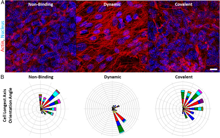
Mammalian cell morphology has been linked to the viscoelastic properties of the adhesion substrate, which is particularly relevant in biological processes such as wound repair and embryonic development where cell spreading and migration are critical. Plastic deformation, degradation, and relaxation of stress are typically coupled in biomaterial systems used to explore these effects, making it unclear which variable drives cell behavior. Here we present a nondegradable polymer architecture that specifically decouples irreversible creep from stress relaxation and modulus. We demonstrate that network plasticity independently controls mesenchymal stem cell spreading through a biphasic relationship dependent on cell-intrinsic forces, and this relationship can be shifted by inhibiting actomyosin contractility. Kinetic Monte Carlo simulations also show strong correlation with experimental cell spreading data as a function of the extracellular matrix (ECM) plasticity. Furthermore, plasticity regulates many ECM adhesion and remodeling genes. Altogether, these findings confirm a key role for matrix plasticity in stem cell biophysics, and we anticipate this will have ramifications in the design of biomaterials to enhance therapeutic applications of stem cells.

Keywords: biomaterials; extracellular matrix; plasticity; stem cell; viscoelasticity.


Conflict of interest statement

The authors declare no competing interest.

Figures

Fig. 1.
Fig. 1.
Schematic depicting how the PEG spacer binding characteristics are proposed to influence hydrogel plasticity in response to cyclical forces. By incorporating pendant PEG to alginate that is either bound covalently, dynamically through Schiff base formation, or simply encapsulated (nonbinding), the irreversible plastic deformations can be controlled with respect to the Young’s modulus and stress relaxation. PEG less constrained to the alginate is proposed to lead to an increase in the force loading of calcium cross-links and thus enhanced irreversible reorganization of the network. In covalent-bound PEG gels, the network responds similarly in response to a singular stress on the network, but upon repeated events, the PEG spacers cannot rearrange to provide optimal plastic behavior. Nonbinding PEG is free-floating and can reorganize in response to stress while the dynamic-binding PEG has intermediate mobility. Individual PEG chains are labeled in the first column to demonstrate chain mobility.
Fig. 2.
Fig. 2.
Modulating the plasticity of alginate hydrogels independently of initial elastic modulus and stress relaxation. (A) Rheological data quantifying the initial Young’s modulus of the various calcium-cross-linked hydrogels. Rheological measurements of five biological replicates. All subsequent experiments were conducted using 10-kDa PEG. (B) Rheological test profile of the stress relaxation assay. (C) The stress relaxation behavior of hydrogels with different incorporation of PEG spacers, analyzed at a strain of 40% with significant overlap. (D) Rheological test profile of the repeated creep assay to probe hydrogel plasticity. (E) Repeated creep studies conducted at 10 Pa for gels with differential incorporation of PEG. (F) The ratio of irreversible to reversible strain due to creep plotted as a function of stress for different PEG incorporation approaches.
Fig. 3.
Fig. 3.
Network plasticity effects on matrix remodeling and adhesion mRNA expression. Cells were encapsulated in alginate hydrogels and incubated for 72 h before mRNA isolation and analysis via NanoString of matrix remodeling and adhesion-associated mRNA. (A) Tree heat map of mRNA expression of five biological replicates of MSCs incubated in each of the three ECMs. (B) Pie chart representing the percentage of DE genes belonging to the most represented family classifications across the three different matrix plasticity conditions. (C) Enrichment analysis of DE genes at different ECM plasticity, measured at 10 Pa of stress. DE was defined as at least a ±2× change and false discovery rate <0.05. (D) Volcano plot of DE genes comparing the nonbinding to a baseline of covalent hydrogels, (E) dynamic to a baseline covalent, and (F) dynamic to baseline nonbinding gels, with statistically-significant DE with P < 0.05 colored in blue. All data are shown with mean ± SD.
Fig. 4.
Fig. 4.
Cell spreading and focal adhesion clustering are enhanced with moderate network plasticity. (A) Representative maximum intensity projection images of MSCs encapsulated within 3.6-kPa (initial modulus) alginate gels with different plasticity. Green represents actin, blue represents the nucleus, and red represents FAK kinase. Confocal images were taken after 3 d in culture. (B) Quantification of the cell aspect ratio, as defined by the 3D rectangular bounding box’s longest side divided by its shortest side. (C) Quantification of the nuclear aspect ratio, as defined by the 3D rectangular bounding box’s longest side divided by its shortest side, showing no significant difference between the conditions. (D) Cellular volume as measured per cell using Imaris Image Analysis software. (E) Representative images of focal adhesions between different binding PEG. (Scale bar, 5 µm.) (F) Histograms of focal adhesion spacing in 3D space, as based on the nearest neighbor distances for k = 4 neighbors. ANOVA analysis resulted in P < 0.001. (G) Cell spreading in gels of Young’s moduli of 1.1, 3.4, and 8 kPa, respectively, as measured by nanoindentation and varying levels of plasticity. *P < 0.1, **P < 0.01, ***P < 0.001, n.s., not significant.
Fig. 5.
Fig. 5.
Relating ECM plasticity to the molecular clutch theory. (A) Schematic describing how viscoplasticity in hydrogels was modeled by incorporating a sliding frictional element into a generalized Maxwell model. (B) Plot of the plasticity of the modified alginate hydrogels (left to right: covalent, dynamic-bis, dynamic, and nonbinding) versus the corresponding experimental cell spreading speeds, with an overlay in red of a KMC simulation of the modified motor-clutch model (n = 50). Spreading of cells in (C) nonbinding, (D) dynamic, and (E) covalent-binding PEG with increasing amounts of blebbistatin. (F) Schematic of the biphasic relationship of matrix plasticity with cell spreading according to molecular clutch theory with the red dots representative of experimental conditions. (G) Interpretation of the impact of adding blebbistatin to cells in various hydrogels; the molecular clutch loading rate is decreased with blebbistatin, shifting the conditions left along the curve. Red dots in F and G represent experimental cell spreading data from CE. YAP nuclear translocation in cells within the (H) nonbinding, (I) dynamic, and (J) covalent-binding PEG with addition of various concentrations of blebbistatin to the culture media. *P < 0.1, **P < 0.01, n.s., not significant.
Fig. 6.
Fig. 6.
Long-term effects of ECM plasticity on stress fiber orientation. (A) Maximum intensity projection of a confocal image of MSCs incubated in hydrogels with different matrix plasticity for 21 d. Actin was stained in red with phalloidin and DAPI stained the nucleus in blue. (Scale bar, 18 μm.) (B) Polar plot histograms of actin fiber orientation angle as analyzed in Imaris of z-stacks. From a range of 0° to 180° with respect to the imaging frame, the actin stress fibers’ longest axis was matched to an angle of best fit.

References

    1. Vining K. H., Mooney D. J., Mechanical forces direct stem cell behaviour in development and regeneration. Nat. Rev. Mol. Cell Biol. 18, 728–742 (2017). - PMC - PubMed
    1. Baker B. M., Chen C. S., Deconstructing the third dimension: How 3D culture microenvironments alter cellular cues. J. Cell Sci. 125, 3015–3024 (2012). - PMC - PubMed
    1. Wang W. Y., Davidson C. D., Lin D., Baker B. M., Actomyosin contractility-dependent matrix stretch and recoil induces rapid cell migration. Nat. Commun. 10, 1186 (2019). - PMC - PubMed
    1. Li Q. et al. ., Extracellular matrix scaffolding guides lumen elongation by inducing anisotropic intercellular mechanical tension. Nat. Cell Biol. 18, 311–318 (2016). - PubMed
    1. Ferreira S. A. et al. ., Neighboring cells override 3D hydrogel matrix cues to drive human MSC quiescence. Biomaterials 176, 13–23 (2018). - PMC - PubMed

Publication types

LinkOut - more resources