

Cryo-EM structures reveal distinct mechanisms of inhibition of the human multidrug transporter ABCB1
- PMID: 33020312
- PMCID: PMC7585025
- DOI: 10.1073/pnas.2010264117
Cryo-EM structures reveal distinct mechanisms of inhibition of the human multidrug transporter ABCB1
Abstract
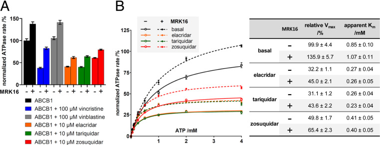
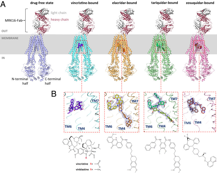
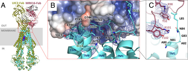
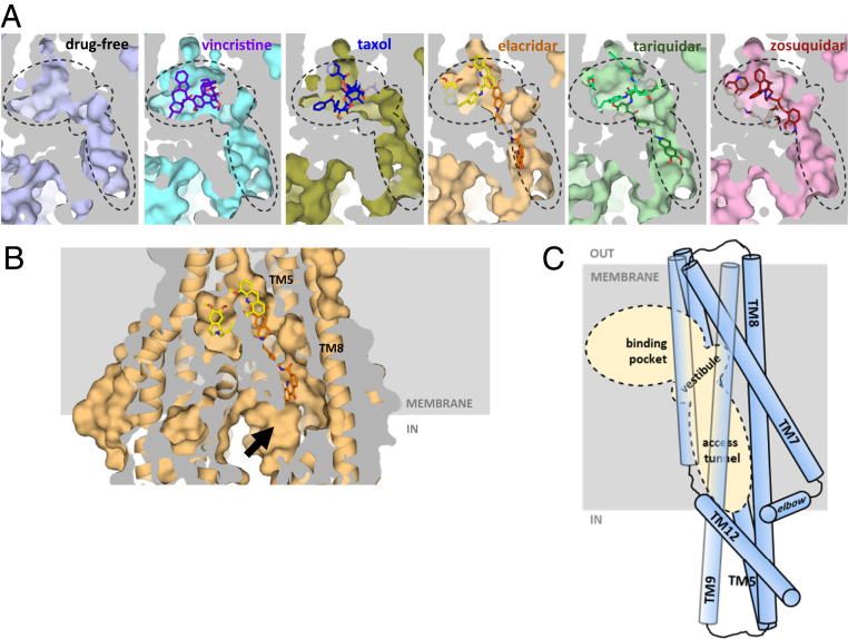
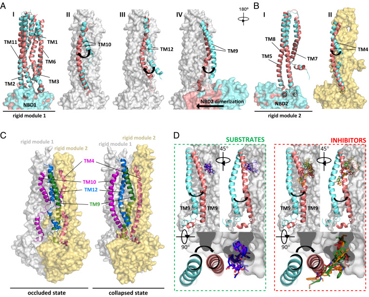
ABCB1 detoxifies cells by exporting diverse xenobiotic compounds, thereby limiting drug disposition and contributing to multidrug resistance in cancer cells. Multiple small-molecule inhibitors and inhibitory antibodies have been developed for therapeutic applications, but the structural basis of their activity is insufficiently understood. We determined cryo-EM structures of nanodisc-reconstituted, human ABCB1 in complex with the Fab fragment of the inhibitory, monoclonal antibody MRK16 and bound to a substrate (the antitumor drug vincristine) or to the potent inhibitors elacridar, tariquidar, or zosuquidar. We found that inhibitors bound in pairs, with one molecule lodged in the central drug-binding pocket and a second extending into a phenylalanine-rich cavity that we termed the "access tunnel." This finding explains how inhibitors can act as substrates at low concentration, but interfere with the early steps of the peristaltic extrusion mechanism at higher concentration. Our structural data will also help the development of more potent and selective ABCB1 inhibitors.
Keywords: ABC transporter; ABCB1; P-glycoprotein; single-particle cryoelectron microscopy; structure.
Conflict of interest statement
The authors declare no competing interest.
Figures
Similar articles
-
Structure of a zosuquidar and UIC2-bound human-mouse chimeric ABCB1.Proc Natl Acad Sci U S A. 2018 Feb 27;115(9):E1973-E1982. doi: 10.1073/pnas.1717044115. Epub 2018 Feb 13. Proc Natl Acad Sci U S A. 2018. PMID: 29440498 Free PMC article.
-
Drug-protein hydrogen bonds govern the inhibition of the ATP hydrolysis of the multidrug transporter P-glycoprotein.Biochem Pharmacol. 2016 Feb 1;101:40-53. doi: 10.1016/j.bcp.2015.12.007. Epub 2015 Dec 11. Biochem Pharmacol. 2016. PMID: 26686578 Free PMC article.
-
Structural insights into binding-site access and ligand recognition by human ABCB1.EMBO J. 2025 Feb;44(4):991-1006. doi: 10.1038/s44318-025-00361-z. Epub 2025 Jan 13. EMBO J. 2025. PMID: 39806099 Free PMC article.
-
Cryo-EM structure of P-glycoprotein bound to triple elacridar inhibitor molecules.Biochem Biophys Res Commun. 2024 May 21;709:149855. doi: 10.1016/j.bbrc.2024.149855. Epub 2024 Mar 28. Biochem Biophys Res Commun. 2024. PMID: 38579618
-
Multidrug transporters: recent insights from cryo-electron microscopy-derived atomic structures and animal models.F1000Res. 2020 Jan 13;9:F1000 Faculty Rev-17. doi: 10.12688/f1000research.21295.1. eCollection 2020. F1000Res. 2020. PMID: 32055397 Free PMC article. Review.
Cited by
-
A Novel 3-meta-Pyridine-1,2,4-oxadiazole Derivative of Glycyrrhetinic Acid as a Safe and Promising Candidate for Overcoming P-Glycoprotein-Mediated Multidrug Resistance in Tumor Cells.ACS Omega. 2023 Dec 12;8(51):48813-48824. doi: 10.1021/acsomega.3c06202. eCollection 2023 Dec 26. ACS Omega. 2023. PMID: 38162726 Free PMC article.
-
Novel betulin derivatives as multidrug reversal agents targeting P-glycoprotein.Sci Rep. 2024 Jan 2;14(1):70. doi: 10.1038/s41598-023-49939-9. Sci Rep. 2024. PMID: 38167542 Free PMC article.
-
Pentagalloyl Glucose-Targeted Inhibition of P-Glycoprotein and Re-Sensitization of Multidrug-Resistant Leukemic Cells (K562/ADR) to Doxorubicin: In Silico and Functional Studies.Pharmaceuticals (Basel). 2023 Aug 22;16(9):1192. doi: 10.3390/ph16091192. Pharmaceuticals (Basel). 2023. PMID: 37765000 Free PMC article.
-
Identification and Empiric Evaluation of New Inhibitors of the Multidrug Transporter P-Glycoprotein (ABCB1).Int J Mol Sci. 2023 Mar 10;24(6):5298. doi: 10.3390/ijms24065298. Int J Mol Sci. 2023. PMID: 36982374 Free PMC article.
-
Structural insight into binding site access and ligand recognition by human ABCB1.bioRxiv [Preprint]. 2024 Aug 12:2024.08.12.607598. doi: 10.1101/2024.08.12.607598. bioRxiv. 2024. Update in: EMBO J. 2025 Feb;44(4):991-1006. doi: 10.1038/s44318-025-00361-z. PMID: 39185192 Free PMC article. Updated. Preprint.
References
Publication types
MeSH terms
Substances
LinkOut - more resources
Full Text Sources
Molecular Biology Databases
