. 2020 Oct 4;10(10):1404.
doi: 10.3390/biom10101404.

Direct Effects of D-Chiro-Inositol on Insulin Signaling and Glucagon Secretion of Pancreatic Alpha Cells

Affiliations

Direct Effects of D-Chiro-Inositol on Insulin Signaling and Glucagon Secretion of Pancreatic Alpha Cells

Agnese Filippello et al. Biomolecules. .

Abstract

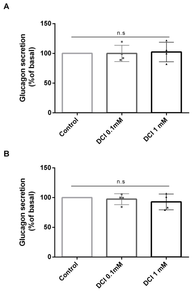
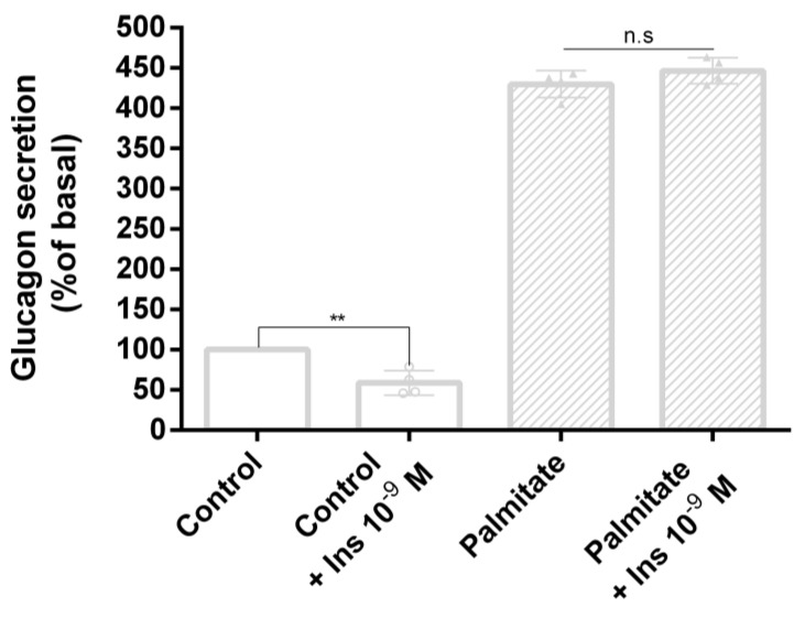
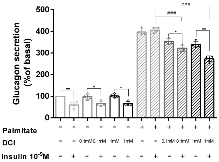
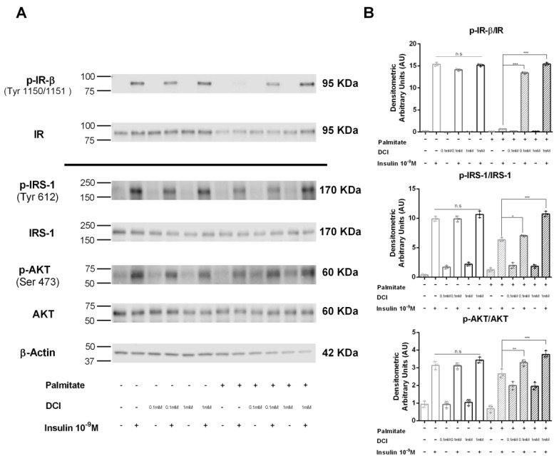
The insulin resistance state of pancreatic α-cells seems to be related to glucagon hypersecretion in type 2 diabetes. Treatment that can improve the insulin sensitivity of α-cells could control glucagon levels in patients with diabetes mellitus. The aim of this study was to investigate the preventive role of D-chiro-inositol (DCI), which has insulin receptor-sensitizer effects on insulin signaling pathways and glucagon secretion in pancreatic α-TC1 clone 6 cells. Cells were chronically treated with palmitate to induce insulin resistance in the presence/absence of DCI. DCI treatment improved the insulin signaling pathway and restored insulin-mediated glucagon suppression in α-TC1-6 cells exposed to palmitate. These results indicate that DCI treatment prevents the insulin resistance of α-TC1-6 cells chronically exposed to palmitate. Our data provide evidence that DCI could be useful to improve the insulin sensitivity of pancreatic α-cells in diabetes treatment.

Keywords: D-chiro-inositol; insulin resistance; lipotoxicity; α-cells.


Conflict of interest statement

The authors declare no conflict of interest.

Figures

Figure 1
Figure 1
Effect of palmitate and D-chiro-inositol (DCI) exposure on cell viability in α-TC1-6 cells. MTT assay in α-TC1-6 cells exposed to increasing concentrations of palmitate or DCI. (A) α-TC1-6 cells exposed to palmitate (0.25, 0.5, 0.75, and 1 mM) for 48 h. (B) α-TC1-6 cells exposed to DCI (0.1, 0.5, 1, and 2 mM) for 48 h. (C) α-TC1-6 cells exposed to DCI (0.1, 0.5, and 1 mM) in the presence of palmitate (0.5 mM) for 48 h. Data are expressed as scatter plots with bar ± standard error of 570 nM absorbance to percentage of control. Experiments were conducted in four biological replicates. One-way ANOVA followed by Bonferroni test (n = 4): * p < 0.05, *** p < 0.001 with respect to controls. n.s., not significant; PA, palmitate; DCI, D-chiro-inositol.
Figure 2
Figure 2
Effect of DCI on glucagon secretion in α-TC1-6 cells. Glucagon secretion in cells treated with DCI (0.1 or 1 mM) for 48 h in control conditions (without palmitate). (A) Acute glucagon levels secreted by α-cells cultured in the presence or absence of DCI; glucagon levels were detected after 48 h of treatment in 2 h cell-conditioned Krebs–Ringer buffer (KRB) (control: 1505 pg/mg protein ± 12 vs. DCI 0.1 mM: 1504 pg/mg protein ± 12 vs. DCI 1 mM: 1539 pg/mg protein ± 13). (B) Chronic glucagon levels, detected directly in Dulbecco’s modified Eagle’s medium (DMEM) at the end of 48 h of treatment in cells cultured in the presence or absence of DCI in control conditions, without palmitate (control: 1825 pg/mg protein ± 14  vs. DCI 0.1 mM: 1774 pg/mg protein ± 9 vs. DCI 1 mM: 1694 pg/mg protein ± 12). Data represent four independent experiments. One-way ANOVA followed by Bonferroni test (n = 4) was used to evaluate statistical significance in DCI treated cells with respect to controls. n.s., not significant; DCI, D-chiro-inositol.
Figure 3
Figure 3
Effect of palmitate on glucagon secretion in α-TC1-6 cells. Insulin-regulated glucagon secretion in control cells and cells treated with palmitate (0.5 mM for 48 h) and/or insulin 10−9 M. Data represent four independent experiments. Student’s t-test was used to evaluate statistical significance in insulin-stimulated cells with respect to unstimulated cells in the absence and presence of palmitate (control: 1405 pg/mg protein ± 13  vs. control + Ins 10–9 M: 829 pg/mg protein ± 11; palmitate: 6036 pg/mg ± 12  vs. palmitate + Ins 10–9 M: 6273 pg/mg protein ± 12 ): ** p < 0.01. n.s., not significant.
Figure 4
Figure 4
Effect of DCI on glucagon secretion in α-TC1-6 cells exposed to palmitate. Acute insulin inhibition of glucagon secretion in control cells and cells treated with 0.1 or 1 mM DCI in the presence or absence of palmitate (0.5 mM for 48 h) and/or insulin 10−9 M. Data represent four independent experiments. Student’s t-test was used to evaluate statistical significance in insulin-stimulated cells with respect to unstimulated cells in the absence and presence of palmitate (control: 1471 pg/mg protein ± 11 vs. control + Ins 10–9 M: 870 pg/mg protein ± 10; palmitate: 5852 pg/mg protein ± 10 vs. palmitate + Ins 10–9 M: 5976 pg/mg protein ± 9); * p < 0.05, ** p < 0.01. One-way ANOVA followed by Bonferroni test (n = 4) was used to evaluate statistical significance in insulin-stimulated cells exposed to increasing DCI concentrations with respect to insulin-stimulated controls in the presence of palmitate. ### p < 0.001. n.s. not significant; DCI, D-chiro-inositol.
Figure 5
Figure 5
Effect of DCI on insulin receptor (IR) phosphorylation and the IRS-1/AKT pathway in α-TC1-6 cells exposed to palmitate. Immunoprecipitation and Western blot analysis for total IR (Tyr1150/1151-β subunit) (p-IR-β) and total IR; Western blot analysis for p-IRS-1 (Tyr612), total IRS-1 (IRS-1), p-AKT (Ser 473), total AKT (AKT), and β-actin: αTC1-6 cells were treated with DCI (0.1 or 1 mM for 48 h) in the presence or absence of palmitate (0.5 mM for 48 h) and acutely stimulated with insulin (10−9 M for 5 min). (A) Representative immunoblot of three independent experiments. (B) Means ± SEM of densitometric analysis. One-way ANOVA followed by Bonferroni test (n = 3) was used to evaluate statistical significance in insulin-stimulated cells treated with increasing concentrations of DCI with respect to insulin-stimulated controls in the absence and presence of palmitate: * p < 0.05, ** p < 0.01, *** p < 0.001. n.s., not significant; DCI, D-chiro-inositol.

References

    1. Muller T.D., Finan B., Bloom S.R., D’Alessio D., Drucker D.J., Flatt P.R., Fritsche A., Gribble F., Grill H.J., Habener J.F., et al. Glucagon-like peptide 1 (GLP-1) Mol. Metab. 2019;30:72–130. doi: 10.1016/j.molmet.2019.09.010. - DOI - PMC - PubMed
    1. Drucker D.J. Mechanisms of Action and Therapeutic Application of Glucagon-like Peptide-1. Cell Metab. 2018;27:740–756. doi: 10.1016/j.cmet.2018.03.001. - DOI - PubMed
    1. Sandoval D.A., D’Alessio D.A. Physiology of proglucagon peptides: Role of glucagon and GLP-1 in health and disease. Physiol. Rev. 2015;95:513–548. doi: 10.1152/physrev.00013.2014. - DOI - PubMed
    1. Unger R.H., Orci L. The essential role of glucagon in the pathogenesis of diabetes mellitus. Lancet. 1975;1:14–16. doi: 10.1016/S0140-6736(75)92375-2. - DOI - PubMed
    1. Cryer P.E. Mechanisms of hypoglycemia-associated autonomic failure in diabetes. N. Engl. J. Med. 2013;369:362–372. doi: 10.1056/NEJMra1215228. - DOI - PubMed