. 2020 Oct 4;21(19):7337.
doi: 10.3390/ijms21197337.

Neuroprotective Effects of Chronic Resveratrol Treatment and Exercise Training in the 3xTg-AD Mouse Model of Alzheimer's Disease

Affiliations

Neuroprotective Effects of Chronic Resveratrol Treatment and Exercise Training in the 3xTg-AD Mouse Model of Alzheimer's Disease

Tom L Broderick et al. Int J Mol Sci. .

Abstract

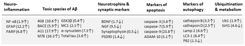
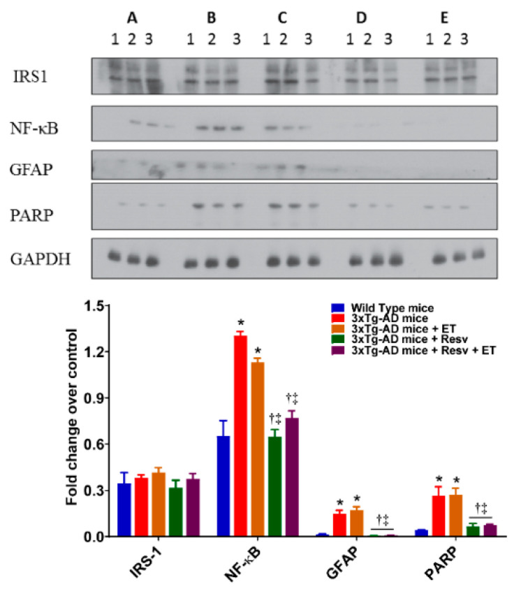
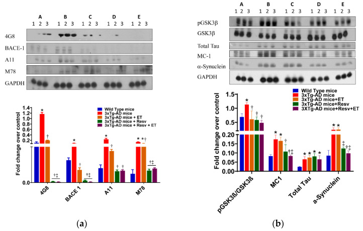
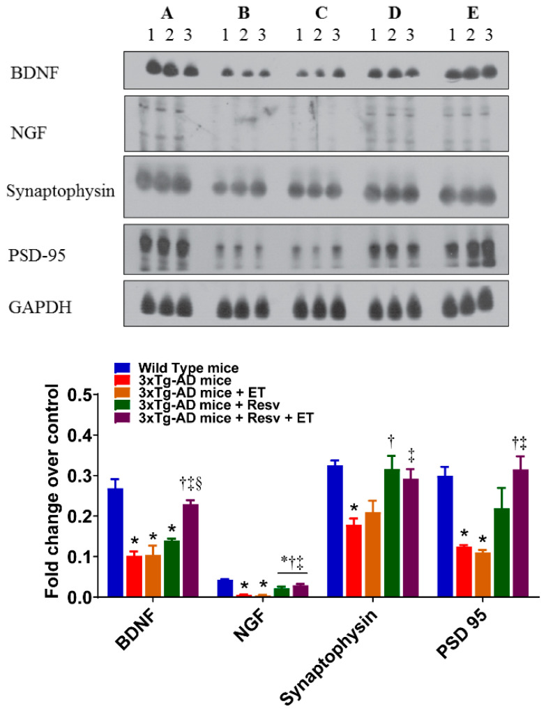
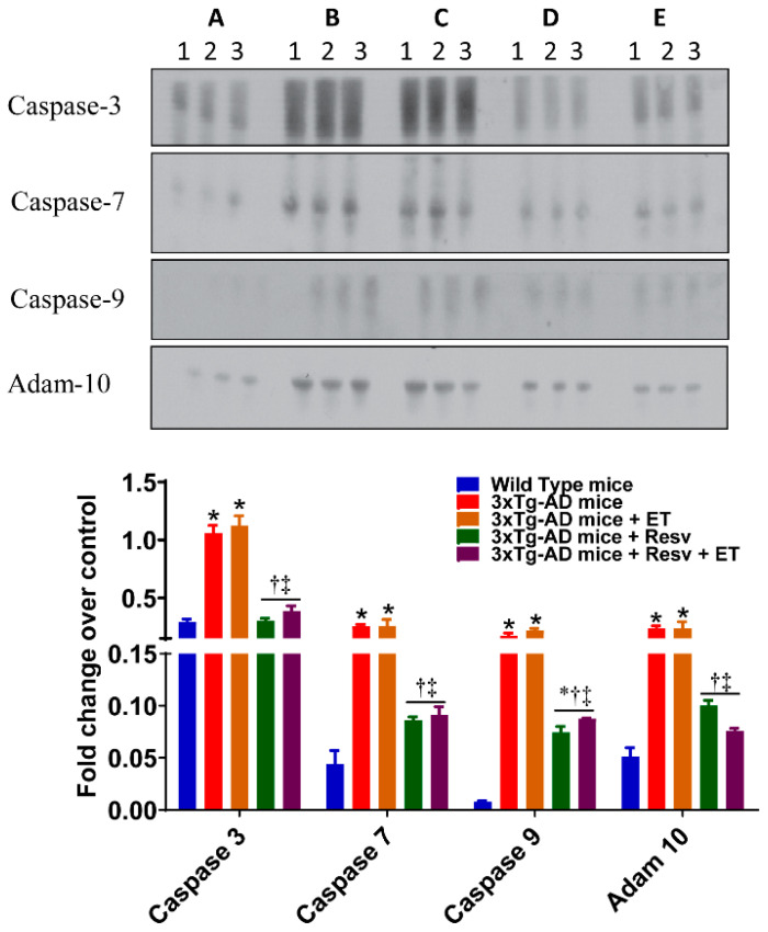
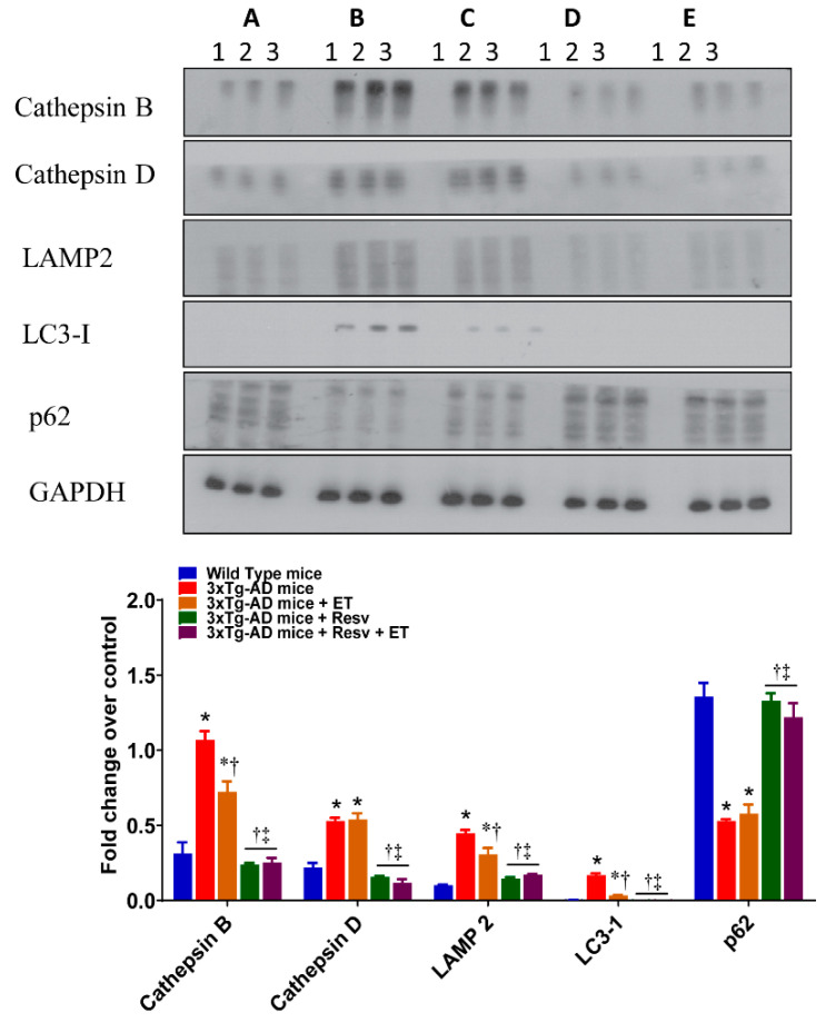
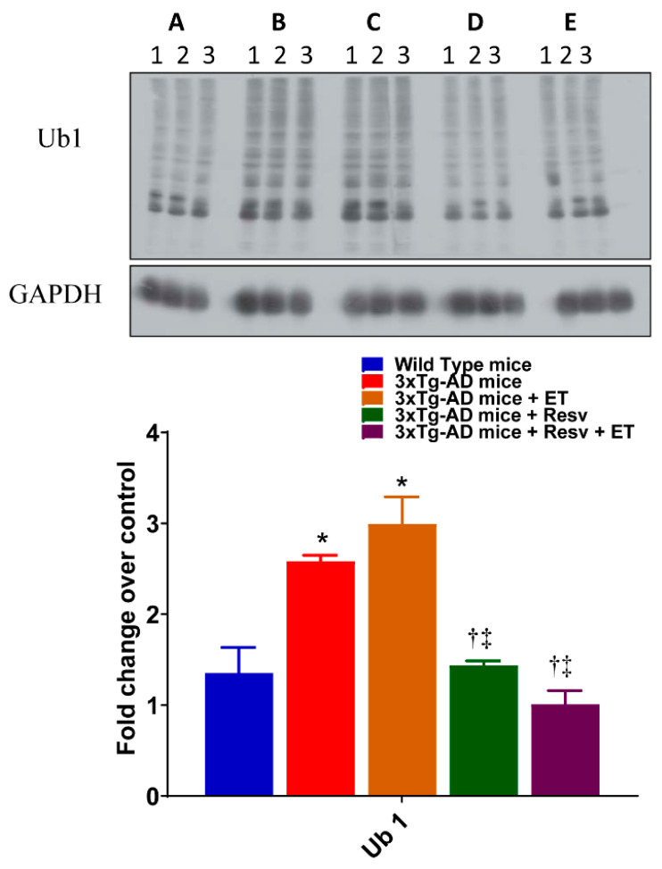
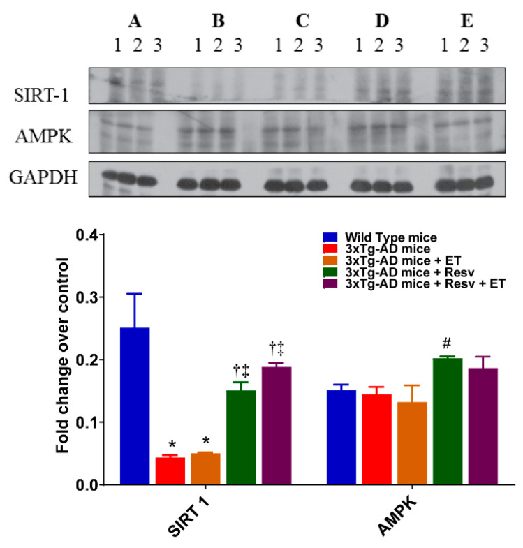
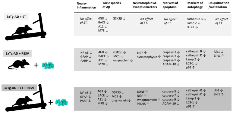
To date, there is no cure or effective treatment for Alzheimer's disease (AD), a chronic neurodegenerative condition that affects memory, language, and behavior. AD is characterized by neuroinflammation, accumulation of brain amyloid-beta (Aβ) oligomers and neurofibrillary tangles, increased neuronal apoptosis, and loss of synaptic function. Promoting regular exercise and a diet containing polyphenols are effective non-pharmacological approaches that prevent the progression of neurodegenerative diseases. In this study, we measured various conformational toxic species of Aβ and markers of inflammation, apoptosis, endolysosomal degradation, and neuroprotection after 5 months of exercise training (ET), resveratrol (Resv) treatment, or combination treatment in the 3xTg-AD mouse model of AD. Our main results indicate that Resv decreased neuroinflammation and accumulation of Aβ oligomers, increased levels of neurotrophins, synaptic markers, silent information regulator, and decreased markers of apoptosis, autophagy, endolysosomal degradation and ubiquitination in the brains of 3xTg-AD mice. ET improved some markers related to neuroprotection, but when combined with Resv treatment, the benefits achieved were as effective as Resv treatment alone. Our results show that the neuroprotective effects of Resv, ET or Resv and ET are associated with reduced toxicity of Aβ oligomers, suppression of neuronal autophagy, decreased apoptosis, and upregulation of key growth-related proteins.

Keywords: Alzheimer’s disease; amyloid-beta; apoptosis; autophagy; brain; exercise; neuroinflammation; neurotrophin; resveratrol.


Conflict of interest statement

The authors declare no conflict of interest.

Figures

Figure 1
Figure 1
Relative changes in the expression of proteins of interest in brain tissue of 3xTg-AD mice. Changes shown as increased (↑) or decreased (↓) in protein expression in brain of 7-month-old 3xTg-AD mice compared to brain from age-matched WT mice. Numbers in parentheses represent the fold change compared to WT mice. Based on the Western blot data, proteins relating to neuroinflammation, toxic species of Aβ, apoptosis, autophagy and ubiquitination were increased whereas protein levels of neurotrophins, synaptic markers and SIRT1 were reduced in the 3xTg-AD mouse brain compared to age-matched WT mice.
Figure 2
Figure 2
The effects of Resv and exercise training (ET) markers of neuroinflammation. Treating 3xTg-AD mice with Resv or Resv with ET decreases the expression of key inflammatory markers. Corresponding densitometry measurements: A, wild-type (WT) mice; B, 3xTg-AD mice; C, 3xTg-AD mice + ET; D, 3xTg-AD mice + Resv; E, 3xTg-AD mice + Resv + ET. NF-κB, nuclear factor-kappa B; GFAP, glial fibrillary acidic protein; PARP, poly (ADP-ribose) polymerase; IRS-1, insulin-receptor substrate. AD, Alzheimer’s disease; ET, exercise training; Resv, resveratrol. Data are presented as mean ± SEM for 3 mice per group. *, compared to WT mice, p < 0.001; , compared to 3xTg-AD mice, p < 0.001; , compared to 3xTg-AD mice + ET, p < 0.05 for NF-κB.
Figure 3
Figure 3
The effects of Resv and ET on conformational toxic species of Aβ. (a) Treating 3xTg-AD mice with Resv or Resv with ET reduces extracellular and intracellular Aβ accumulation in brain. Aβ content, oligomers of Aβ and intracellular Aβ detected by A11 and M78 antibody. (b) Protein content of pGSK3-β was reduced in 3xTg-AD mice treated with Resv, ET, and Resv with ET. Tau oligomers in brain detected by using the MC1 antibody were also reduced with treatment with Resv or Resv with ET but with no change in total tau. Misfolded protein expression levels using α-synuclein antibody were reduced in 3xTg-AD mice treated with Resv or Resv with ET. Corresponding densitometry measurements: A, WT mice; B, 3xTg-AD mice; C, 3xTg-AD mice + ET; D, 3xTg-AD mice Resv; E, 3xTg-AD mice + Resv + ET. BACE1, beta-secretase enzyme 1; pGSK3β, glycogen synthase kinase beta (p = phosphorylated). AD, Alzheimer’s disease; ET, exercise training; Resv, resveratrol. Data are presented as mean ± SEM for 3 mice per group. *, compared to WT mice, p < 0.01; , compared to 3xTg-AD mice, p < 0.01; , compared to 3xTg-AD mice + ET, p < 0.01.
Figure 4
Figure 4
The effects of Resv and ET on the expression of neurotrophins and synaptic markers. Treatment with Resv or Resv with ET increased the expression of neurotrophins and synaptic markers. Representative immunoblots show significant increases in expression of the neurotrophins BDNF, NGF and synaptic markers synaptophysin and PSD-95 in 3xTg-AD mice treated with Resv or Resv with ET. Corresponding densitometry measurements: A, WT mice; B, 3xTg-AD mice; C, 3xTg-AD mice + ET; D, 3xTg-AD mice + Resv; E, 3xTg-AD mice + Resv + ET. BDNF, brain-derived neurotropic factor; NGF, nerve growth factor; PSD95, postsynaptic density 95. AD, Alzheimer’s disease; ET, exercise training; Resv, resveratrol. Data are presented as mean ± SEM for 3 mice per group. *, compared to WT mice, p < 0.05; , compared to 3xTg-AD mice, p < 0.05; , compared to 3xTg-AD mice + ET, p < 0.05; §, compared to 3xTg-AD mice + Resv.
Figure 5
Figure 5
The effects of Resv and ET on the expression of markers of apoptosis. Treatment with Resv or Resv with ET decreased the expression of markers of apoptosis. Representative immunoblots show significant decreases in the expression of caspase 3, 7, 9, and Adam 10. Corresponding densitometry measurements: A, WT mice; B, 3xTg-AD mice; C, 3xTg-AD mice + ET; D, 3xTg-AD mice + Resv; E, 3xTg-AD mice + Resv + ET. Adam 10, disintegrin and metallopeptidase domain-containing protein 10. AD, Alzheimer’s disease; ET, exercise training; Resv, resveratrol. Data are presented as mean ± SEM for 3 mice per group. *, compared to WT mice, p < 0.05; , compared to 3xTg-AD mice, p < 0.05; , compared to 3xTg-AD mice + ET, p < 0.05.
Figure 6
Figure 6
The effects of Resv and ET on the expression of markers of autophagy. Treatment with Resv or Resv with ET decreased protein expression of key autophagy markers. Autophagy was expressed using LC3-1 and Cathepsin B, Cathepsin D, and LAMP2 were measured as endolysosomal markers. Representative immunoblots show significant reductions in the expression of all markers with ET, Resv, and Resv with ET. The significant decrease in the expression p62 in brains of 3xTg-AD mice was prevented with Resv or Resv with ET. Corresponding densitometry measurements: A, WT mice; B, 3xTg-AD mice; C, 3xTg-AD mice+ET; D, 3xTg-AD mice + Resv; E, 3xTg-AD mice + Resv + ET. LAMP2, lysosomal-associated membrane protein; LC3-1, microtubule-associated protein 1 light chain 3 beta; p62, sequestosome 1/ubiquitin-binding protein. AD, Alzheimer’s disease; ET, exercise training; Resv, resveratrol. Data are presented as mean ± SEM for 3 mice per group. *, compared to WT mice, p < 0.05; , compared to 3xTg-AD mice, p < 0.05; , compared to 3xTg-AD mice + ET, p < 0.05.
Figure 7
Figure 7
The effects of Resv and ET on protein ubiquitination. Treatment with Resv or Resv with ET decreased protein ubiquitination, expressed as Ub1. Corresponding densitometry measurements: A, WT mice; B, 3xTg-AD mice; C, 3xTg-AD mice + ET; D, 3xTg-AD mice + Resv; E, 3xTg-AD mice + Resv + ET. AD, Alzheimer’s disease; ET, exercise training; Resv, resveratrol. Data presented as mean ± SD for 3 mice per group. *, compared to WT mice, p < 0.05; , compared to 3xTg-AD mice, p < 0.05; , compared to 3xTg-AD mice + ET, p < 0.05.
Figure 8
Figure 8
The effects of Resv and ET on protein expression of SIRT1 and AMPK. Representative immunoblots show a significant decrease in the expression level of SIRT1 in 3xTg-AD mice. Treatment with Resv or Resv with ET increased the expression SIRT1. Expression levels of AMPK were not significantly different between groups. Corresponding densitometry measurements: A, WT mice; B, 3xTg-AD mice; C, 3xTg-AD mice + ET; D, 3xTg-AD mice + Resv; E, 3xTg-AD mice + Resv + ET. SIRT1, silent information regulator 1; AMPK, AMP-activated protein kinase. AD, Alzheimer’s disease; ET, exercise training; Resv, resveratrol. Data presented as mean ± SEM for 3 mice per group. *, compared to WT mice, p < 0.05; , compared to 3xTg-AD mice, p < 0.05; , compared to 3xTg-AD mice + ET, p < 0.05; #, p = 0.0578 compared to WT mice.
Figure 9
Figure 9
Summary of the effects of Resv and ET on proteins of interest. Changes are expressed as increased (↑) or decreased (↓) based on the Western blot data compared to brain tissue obtained from 3xTg-AD mice serving as controls (Figure 1). The greatest protection was afforded by treatment with Resv alone or by Resv combined with ET.

Similar articles

Cited by

References

    1. Sperling R.A., Dickerson B.C., Pihlajamaki M., Vannini P., LaViolette P.S., Vitolo O.V., Hedden T., Becker J.A., Rentz D.M., Selkoe D.J., et al. Functional Alterations in Memory Networks in Early Alzheimer’s Disease. Neuromol. Med. 2010;12:27–43. doi: 10.1007/s12017-009-8109-7. - DOI - PMC - PubMed
    1. World Alzheimer Report 2018. [(accessed on 18 July 2020)]; Available online: https://www.alz.co.uk/research/world-report-2018.
    1. Harris M.E., Hensley K., Butterfield D.A., Leedle R.A., Carney J.M. Direct evidence of oxidative injury produced by the Alzheimer’s beta-amyloid peptide (1-40) in cultured hippocampal neurons. Exp. Neurol. 1995;131:193–202. doi: 10.1016/0014-4886(95)90041-1. - DOI - PubMed
    1. Eckert A., Hauptmann S., Scherping I., Rhein V., Müller-Spahn F., Götz J., Müller W.E. Soluble beta-amyloid leads to mitochondrial defects in amyloid precursor protein and tau transgenic mice. Neurodegener. Dis. 2008;5:157–159. doi: 10.1159/000113689. - DOI - PubMed
    1. Kolarova M., García-Sierra F., Bartos A., Ricny J., Ripova D. Structure and pathology of tau protein in Alzheimer disease. Int. J. Alzheimers Dis. 2012;2012:731526. doi: 10.1155/2012/731526. - DOI - PMC - PubMed