. 2020 Dec 1:878:114733.
doi: 10.1016/j.jelechem.2020.114733. Epub 2020 Sep 30.

Charge transfer reaction mechanisms of epoxyketone and boronated peptides at glassy carbon and boron doped diamond electrodes

Affiliations

Charge transfer reaction mechanisms of epoxyketone and boronated peptides at glassy carbon and boron doped diamond electrodes

Catarina Sofia Henriques de Jesus et al. J Electroanal Chem (Lausanne). .

Abstract

The ubiquitin-proteasome system regulates the level of proteins within cells through controlled proteolysis. In some diseases, the system function is dysregulated turning the ubiquitin-proteasome complex into a target for drug development. The redox behavior of proteasome inhibitors, epoxyketone and boronated peptides carfilzomib, oprozomib and delanzomib was investigated by voltammetric methods using glassy carbon and boron doped diamond electrodes. It was showed that the oxidation of epoxyketone peptides carfilzomib and oprozomib occurred in one step at glassy carbon electrode surface while at boron doped diamond two consecutive charge transfer reactions due to different adsorption orientation at the electrode surface were observed. The moieties of these peptides, involved in the oxidation process, were morpholine for carfilzomib and thiazole for oprozomib. For the boronated peptide delanzomib, two irreversible and independent redox processes, oxidation at +0.80 V and reduction at -1.40 V were identified in neutral media at both electrodes. The oxidation reaction occurred at the amino group close to the pyridine moiety of delanzomib with the transfer of one electron and one proton whereas the reduction process takes place at pyridine ring in a two-electrons two-protons mechanism. Redox mechanisms were proposed and the implications on the proteasome inhibition discussed.

Keywords: Boron doped diamond; Carfilzomib; Delanzomib; Glassy carbon; Oprozomib; Redox mechanism.


Conflict of interest statement

The authors declare that they have no known competing financial interests or personal relationships that could have appeared to influence the work reported in this paper.

Figures

Unlabelled Image
Graphical abstract
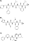
Scheme 1
Scheme 1
Chemical structures of A) carfilzomib, B) oprozomib and C) delanzomib.
Fig. 1
Fig. 1
Cyclic voltammogram with A) GCE and B) BDDE in solution of 25 μM carfilzomib in 0.1 M phosphate buffer pH = 7.0 at v = 100 mV s−1; (—) 1st, (…) 2nd and (…) 3rd scans.
Fig. 2
Fig. 2
1) DP voltammogram in solution of 25 μM carfilzomib function of pH of the supporting electrolyte with A) GCE and B) BDDE. 2) Plots of the variation of (○,□) Ipa and (●,■) Epa of carfilzomib vs. pH. The red curves correspond to voltammograms recorded in solutions of methyl-morpholine. (For interpretation of the references to colour in this figure legend, the reader is referred to the web version of this article.)
Fig. 3
Fig. 3
SW voltammograms with A) GCE and B) BDDE in solution of 25 μM carfilzomib in 0.1 M phosphate buffer pH = 7.0 at v = 100 mV s−1.
Fig. 4
Fig. 4
Cyclic voltammogram with BDDE in solution of: A) different concentrations and B) 100 μM oprozomib (—) 1st, (…) 2nd and (…) 3rd scans, in 0.1 M phosphate buffer pH = 7.0 at v = 100 mV s−1.
Fig. 5
Fig. 5
A, B) DP and C) SW voltammograms with A) GCE and B,C) BDDE in solutions of A) 75 μM oprozomib in electrolytes with different pH values, B) different concentrations of oprozomib in 0.1 M phosphate buffer pH = 7.0, and C) 75 μM oprozomib in 0.1 M phosphate buffer pH = 7.0. For SWV, It, Ib and If represents the total, backward and forward currents.
Fig. 6
Fig. 6
Cyclic voltammograms obtained with GCE in solutions of 200 μM delanzomib in 0.1 M phosphate buffer pH = 7.0 between potential limits of: A) + 1.40 and − 1.50 V; B) 0 and + 1.40 V; and C) 0 and − 1.60 V; v = 100 mV s−1; (—) 1st, (…) 2nd and (…) 3rd scans.
Fig. 7
Fig. 7
DP voltammograms obtained with GCE in solution containing different concentration of delanzomib in 0.1 M phosphate buffer pH = 7.0: A) (—) 1st, (…) 2nd and (…) 3rd anodic scan for 200 μM; B) first scan for (—) 1, (…) 10 and (…) 100 μM and C) (—) 1st, () 2nd and (…) 3rd cathodic scan in 200 μM delanzomib.
Fig. 8
Fig. 8
DP voltammograms recorded at GCE in 100 μM morpholine and methyl-morpholine, and in 10 μM 2-aminothiazole and 2-methylthiazole in 0.1 M phosphate buffer pH = 7.0.
Scheme 2
Scheme 2
– Proposed redox mechanisms for A) carfilzomib, B) oprozomib and C) delanzomib.

References

    1. Heinemeyer W., Ramos P.C., Dohmen R.J. Ubiquitin-proteasome system. Cell. Mol. Life Sci. 2004;61:1562–1578. doi: 10.1007/s00018-004-4130-z. - DOI - PMC - PubMed
    1. Ciechanover A. Intracellular protein degradation: from a vague idea thru the lysosome and the ubiquitin–proteasome system and onto human diseases and drug targetingNobel lecture 2004, © the Nobel foundation 2004. Cell Death Differ. 2005;12:1178–1190. doi: 10.1038/sj.cdd.4401692. - DOI - PubMed
    1. Cao B., Mao X. The ubiquitin-proteasomal system is critical for multiple myeloma: implications in drug discovery. Am. J. Blood Res. 2011;1:46–56. - PMC - PubMed
    1. Moreau P., Richardson P.G., Cavo M., Orlowski R.Z., San Miguel J.F., Palumbo A., Harousseau J.-L. Proteasome inhibitors in multiple myeloma: 10 years later. Blood. 2012;120:947–959. doi: 10.1182/blood-2012-04-403733. - DOI - PMC - PubMed
    1. Grosicki S., Barchnicka A., Jurczyszyn A., Grosicka A. Bortezomib for the treatment of multiple myeloma. Expert. Rev. Hematol. 2014;7:173–185. doi: 10.1586/17474086.2014.899144. - DOI - PubMed

LinkOut - more resources