

Apolipoprotein A-I in mouse cerebrospinal fluid derives from the liver and intestine via plasma high-density lipoproteins assembled by ABCA1 and LCAT
- PMID: 33020907
- PMCID: PMC7987658
- DOI: 10.1002/1873-3468.13950
Apolipoprotein A-I in mouse cerebrospinal fluid derives from the liver and intestine via plasma high-density lipoproteins assembled by ABCA1 and LCAT
Abstract
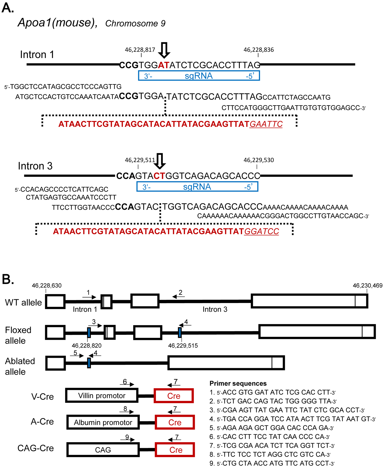
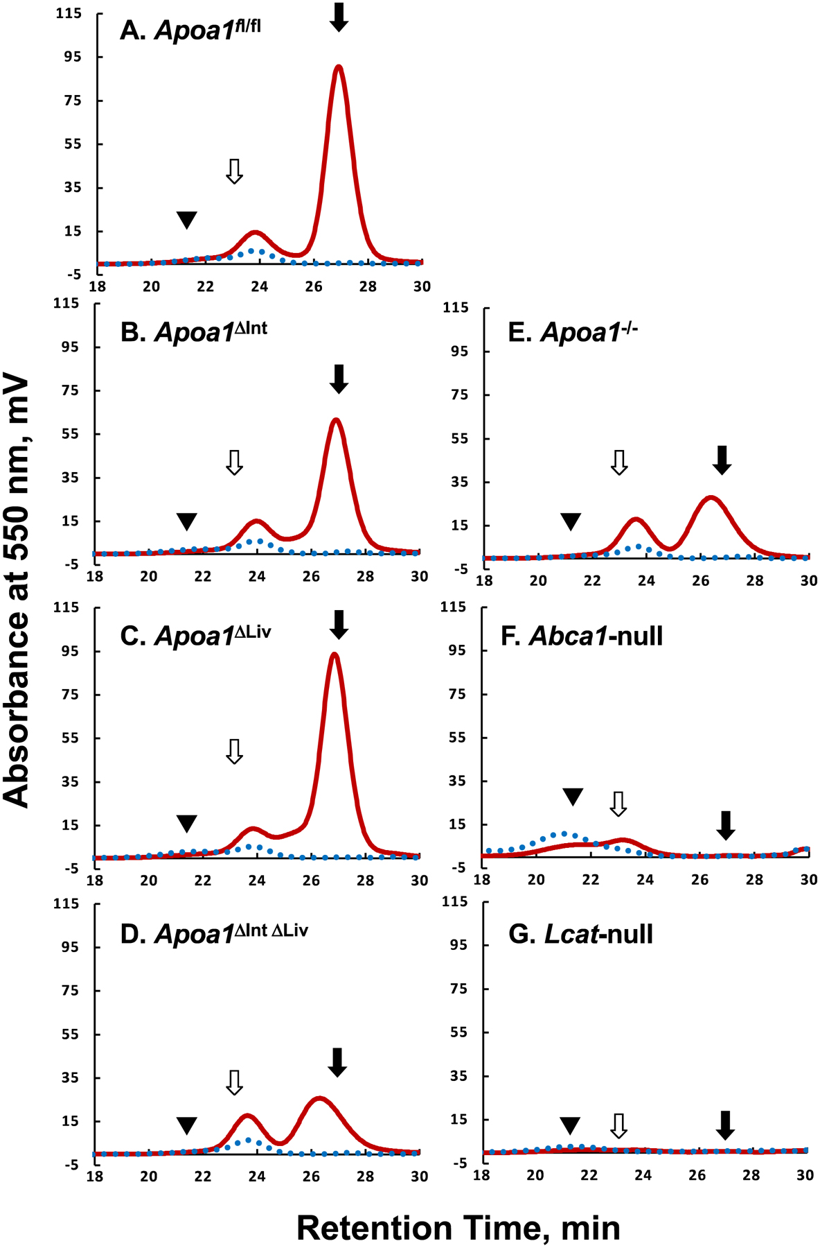
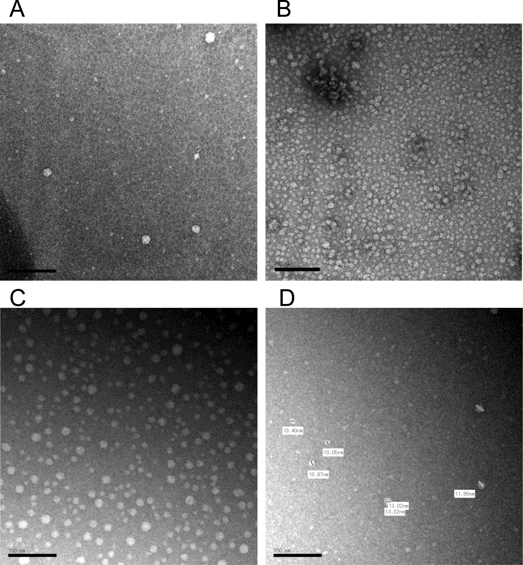
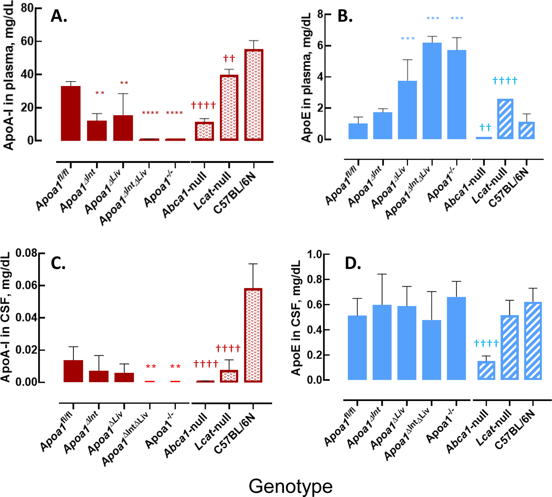
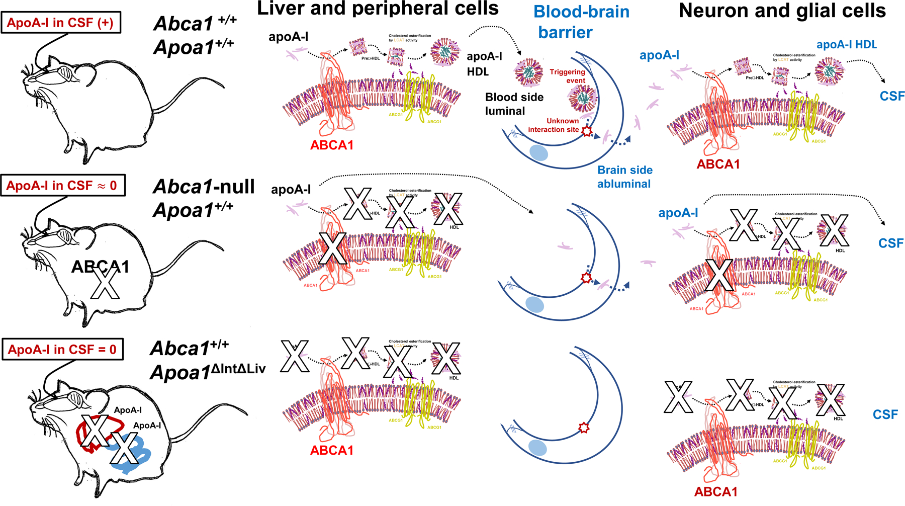
Apolipoprotein (apo) A-I, the major structural protein of high-density lipoprotein (HDL), is present in human and mouse cerebrospinal fluid (CSF) despite its lack of expression in brain cells. To identify the origin of apoA-I in CSF, we generated intestine-specific and liver-specific Apoa1 knockout mice (Apoa1ΔInt and Apoa1Δliv mice, respectively). Lipoprotein profiles of Apoa1ΔInt and Apoa1ΔLiv mice resembled those of control littermates, whereas knockout of Apoa1 in both intestine and liver (Apoa1ΔIntΔLiv ) resulted in a 60-percent decrease in HDL-cholesterol levels, thus strongly mimicking the Apoa1-/- mice. Immunoassays revealed that mouse apoA-I was not present in the CSF of the Apoa1ΔIntΔLiv mice. Furthermore, apoA-I levels in CSF were highly correlated with plasma spherical HDL levels, which were regulated by ABCA1 and LCAT. Collectively, these results suggest that apoA-I protein in CSF originates in liver and small intestine and is taken up from the plasma.
Keywords: apoA-I; cerebrospinal fluid; intestine; liver; mice.
© 2020 Federation of European Biochemical Societies.
Conflict of interest statement
Disclosure of conflicts of interest:
The authors declare no financial or other conflicts of interest.
Figures

Similar articles
-
Naturally occurring and bioengineered apoA-I mutations that inhibit the conversion of discoidal to spherical HDL: the abnormal HDL phenotypes can be corrected by treatment with LCAT.Biochem J. 2007 Aug 15;406(1):167-74. doi: 10.1042/BJ20070296. Biochem J. 2007. PMID: 17506726 Free PMC article.
-
LCAT can rescue the abnormal phenotype produced by the natural ApoA-I mutations (Leu141Arg)Pisa and (Leu159Arg)FIN.Biochemistry. 2007 Sep 18;46(37):10713-21. doi: 10.1021/bi7003203. Epub 2007 Aug 21. Biochemistry. 2007. PMID: 17711302
-
Substitutions of glutamate 110 and 111 in the middle helix 4 of human apolipoprotein A-I (apoA-I) by alanine affect the structure and in vitro functions of apoA-I and induce severe hypertriglyceridemia in apoA-I-deficient mice.Biochemistry. 2004 Aug 17;43(32):10442-57. doi: 10.1021/bi049782p. Biochemistry. 2004. PMID: 15301543
-
Role of apoA-I, ABCA1, LCAT, and SR-BI in the biogenesis of HDL.J Mol Med (Berl). 2006 Apr;84(4):276-94. doi: 10.1007/s00109-005-0030-4. Epub 2006 Feb 25. J Mol Med (Berl). 2006. PMID: 16501936 Review.
-
Discrete roles of apoA-I and apoE in the biogenesis of HDL species: lessons learned from gene transfer studies in different mouse models.Ann Med. 2008;40 Suppl 1:14-28. doi: 10.1080/07853890701687219. Ann Med. 2008. PMID: 18246469 Review.
Cited by
-
Apolipoprotein A (ApoA) in Neurological Disorders: Connections and Insights.Int J Mol Sci. 2025 Aug 16;26(16):7908. doi: 10.3390/ijms26167908. Int J Mol Sci. 2025. PMID: 40869228 Free PMC article. Review.
-
An Improved Method for Sampling and Quantitative Protein Analytics of Cerebrospinal Fluid of Individual Mice.Mol Cell Proteomics. 2025 May;24(5):100958. doi: 10.1016/j.mcpro.2025.100958. Epub 2025 Mar 27. Mol Cell Proteomics. 2025. PMID: 40157722 Free PMC article.
-
Elucidation of physico-chemical principles of high-density lipoprotein-small RNA binding interactions.J Biol Chem. 2022 Jun;298(6):101952. doi: 10.1016/j.jbc.2022.101952. Epub 2022 Apr 18. J Biol Chem. 2022. PMID: 35447119 Free PMC article.
-
Lipoprotein Particles in Cerebrospinal Fluid.Arterioscler Thromb Vasc Biol. 2024 May;44(5):1042-1052. doi: 10.1161/ATVBAHA.123.318284. Epub 2024 Mar 28. Arterioscler Thromb Vasc Biol. 2024. PMID: 38545782 Free PMC article. Review.
-
Cerebrospinal Fluid Proteomic Profiles in Patients with Postherpetic Neuralgia.J Proteome Res. 2023 Dec 1;22(12):3879-3892. doi: 10.1021/acs.jproteome.3c00547. Epub 2023 Nov 15. J Proteome Res. 2023. PMID: 37966014 Free PMC article.
References
-
- Gordon T, Castelli WP, Hjortland MC, Kannel WB and Dawber TR (1977). High density lipoprotein as a protective factor against coronary heart disease. The Framingham Study. Am J Med 62, 707–14. - PubMed
Publication types
MeSH terms
Substances
Grants and funding
LinkOut - more resources
Full Text Sources
Molecular Biology Databases
Miscellaneous
