Review
. 2020 Oct;150(1):47-56.
doi: 10.1007/s11060-020-03639-w. Epub 2020 Oct 6.

Atypical teratoid rhabdoid tumor: molecular insights and translation to novel therapeutics

Affiliations
Review

Atypical teratoid rhabdoid tumor: molecular insights and translation to novel therapeutics

Cody L Nesvick et al. J Neurooncol. 2020 Oct.

Abstract

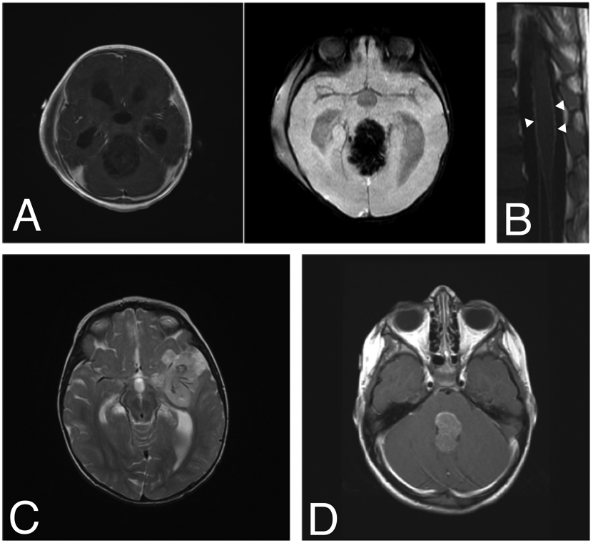
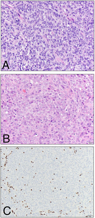
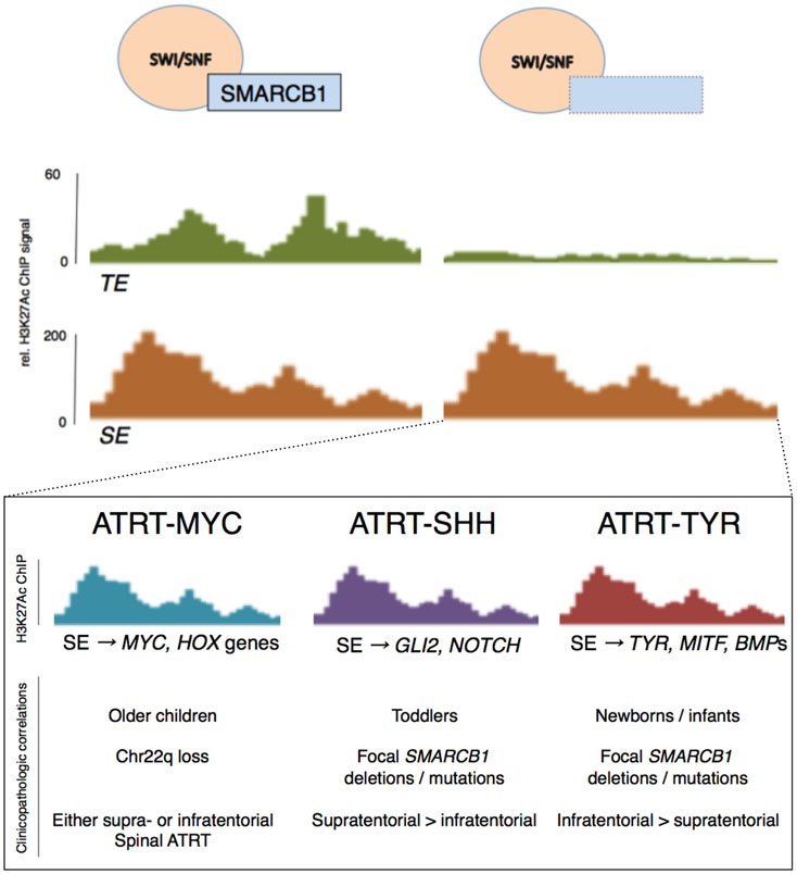
Introduction: Atypical teratoid rhabdoid tumor (ATRT) is a rare, often lethal brain tumor of childhood characterized by a complex epigenetic landscape amongst a simple genetic background. Recent molecular studies have defined key biologic events that contribute to tumorigenesis and molecular subtypes of ATRT.

Methods: Seminal studies on ATRT are reviewed with an emphasis on molecular pathogenesis and its relevance to novel therapeutics.

Results: In this review, we summarize the key clinicopathologic and molecular features of ATRT, completed and ongoing clinical trials and outline the translational potential of novel insights into the molecular pathogenesis of this tumor.

Conclusions: SMARCB1 loss is the key genetic event in ATRT pathogenesis that leads to widespread epigenetic dysregulation and loss of lineage-specific enhancers. Current work is defining subtype-specific treatments that target underlying molecular derangements that drive tumorigenesis.

Keywords: ATRT; Atypical teratoid rhabdoid tumor; Chromatin remodeling; Epigenetics; SWI/SNF complex.


Conflict of interest statement

Conflicts of interest: The authors have no conflicts of interest to declare.

Figures

Figure 1.
Figure 1.. A spectrum of clinical imaging findings of ATRT.
ATRT should be considered in any young child with imaging features of a highly aggressive brain tumor. A) A 9-month-old boy presented with vomiting followed quickly by coma. MRI demonstrated a 6-cm pineal-region mass with minimal, heterogeneous enhancement and abundant vascularity on susceptibility-weighted imaging. B) Diffuse leptomeningeal enhancement of the spinal cord and cauda equina were noted at the time of diagnosis (arrowheads). He underwent a gross total resection (GTR) via suboccipital craniotomy followed by ACNS0333 chemotherapy, stem cell rescue and proton beam radiotherapy with adjuvant craniospinal radiation (CSI). He currently has no evidence of disease three and a half years after diagnosis. C) A 13-month-old girl presented with coma and was found to have a 4-cm left frontotemporal, minimally enhancing mass encasing several large feeding blood vessels (T2-weighted image with contrast shown). She underwent GTR followed by adjuvant chemotherapy using the ACNS0333 protocol, stem-cell transplant and CSI. She is currently seven years since diagnosis and remains disease-free. D) A 4-year-old girl presented with nausea, vomiting and lethargy. MRI demonstrated a 3-cm, homogeneously enhancing fourth ventricular mass (T1-weighted image with contrast shown). She underwent near-total resection via suboccipital craniotomy, adjuvant chemotherapy with the SJMB-06 protocol and CSI. She is free of disease six years post-operatively.
Figure 2.
Figure 2.. Histological features of ATRT.
A) The hypercellular tumor is usually predominantly composed of primitive-appearing cells with scant cytoplasm and hyperchromatic nuclei. Mitotic figures, apoptotic bodies and necrosis may all be readily identified. B) A subset of cells may show abundant, globular eosinophilic cytoplasm, reminiscent of rhabdoid cells. C) SMARCB1 (INI1 / BAF47 / SNF5) immunohistochemistry shows uniform loss of expression in the tumor cells, while expression is retained in endothelial nuclei serving as positive internal controls. (A, B – Hematoxylin and Eosin stain, 400x magnification; C – BAF47 clone (BD Biosciences), 200x magnification).
Figure 3.
Figure 3.. Enhancer landscape in ATRT and specificity to molecular subtypes.
Loss of SMARCB1 results in selective loss of typical enhancers (TEs) involved in cell lineage determination via residual activity of the SWI/SNF complex, loss of histone acetylase activity and increased relative PRC2/EZH2 activity. However, super enhancer (SE) activity is selectively retained at genes that contribute to cell proliferation and immortality in one of three specific patterns. These three molecular subgroups of ATRT are relevant to developing logical, targeted therapies and potentially prognostication.

References

    1. Louis DN, Perry A, Reifenberger G, et al. (2016) The 2016 World Health Organization Classification of Tumors of the Central Nervous System: a summary. Acta Neuropathol 131:803–20. 10.1007/s00401-016-1545-1 - DOI - PubMed
    1. Ellison D, Love S (2013) Neuropathology: A Reference Text of CNS Pathology, 3rd ed. Mosby Ltd
    1. Sévenet N, Sheridan E, Amram D, et al. (1999) Constitutional mutations of the hSNF5/INI1 gene predispose to a variety of cancers. Am J Hum Genet 65:1342–1348. 10.1086/302639 - DOI - PMC - PubMed
    1. Biegel JA, Tan L, Zhang F, et al. (2002) Alterations of the hSNF5/INI1 gene in central nervous system atypical teratoid/rhabdoid tumors and renal and extrarenal rhabdoid tumors. Clin Cancer Res 8:3461–3467 - PubMed
    1. Versteege I, Sévenet N, Lange J, et al. (1998) Truncating mutations of hSNF5/INI1 in aggressive paediatric cancer. Nature 394:203–6. 10.1038/28212 - DOI - PubMed

Supplementary concepts

LinkOut - more resources