Review
. 2020 Nov 1;93(1115):20200172.
doi: 10.1259/bjr.20200172. Epub 2020 Oct 6.

The 20th Gray lecture 2019: health and heavy ions

Affiliations
Review

The 20th Gray lecture 2019: health and heavy ions

Eleanor A Blakely. Br J Radiol. .

Abstract

Objective: Particle radiobiology has contributed new understanding of radiation safety and underlying mechanisms of action to radiation oncology for the treatment of cancer, and to planning of radiation protection for space travel. This manuscript will highlight the significance of precise physical and biologically effective dosimetry to this translational research for the benefit of human health.This review provides a brief snapshot of the evolving scientific basis for, and the complex current global status, and remaining challenges of hadron therapy for the treatment of cancer. The need for particle radiobiology for risk planning in return missions to the Moon, and exploratory deep-space missions to Mars and beyond are also discussed.

Methods: Key lessons learned are summarized from an impressive collective literature published by an international cadre of multidisciplinary experts in particle physics, radiation chemistry, medical physics of imaging and treatment planning, molecular, cellular, tissue radiobiology, biology of microgravity and other stressors, theoretical modeling of biophysical data, and clinical results with accelerator-produced particle beams.

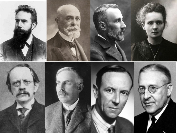
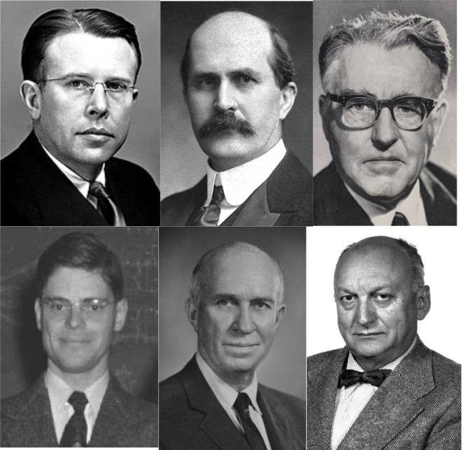
Results: Research pioneers, many of whom were Nobel laureates, led the world in the discovery of ionizing radiations originating from the Earth and the Cosmos. Six radiation pioneers led the way to hadron therapy and the study of charged particles encountered in outer space travel. Worldwide about 250,000 patients have been treated for cancer, or other lesions such as arteriovenous malformations in the brain between 1954 and 2019 with charged particle radiotherapy, also known as hadron therapy. The majority of these patients (213,000) were treated with proton beams, but approximately 32,000 were treated with carbon ion radiotherapy. There are 3500 patients who have been treated with helium, pions, neon or other ions. There are currently 82 facilities operating to provide ion beam clinical treatments. Of these, only 13 facilities located in Asia and Europe are providing carbon ion beams for preclinical, clinical, and space research. There are also numerous particle physics accelerators worldwide capable of producing ion beams for research, but not currently focused on treating patients with ion beam therapy but are potentially available for preclinical and space research. Approximately, more than 550 individuals have traveled into Lower Earth Orbit (LEO) and beyond and returned to Earth.

Conclusion: Charged particle therapy with controlled beams of protons and carbon ions have significantly impacted targeted cancer therapy, eradicated tumors while sparing normal tissue toxicities, and reduced human suffering. These modalities still require further optimization and technical refinements to reduce cost but should be made available to everyone in need worldwide. The exploration of our Universe in space travel poses the potential risk of exposure to uncontrolled charged particles. However, approaches to shield and provide countermeasures to these potential radiation hazards in LEO have allowed an amazing number of discoveries currently without significant life-threatening medical consequences. More basic research with components of the Galactic Cosmic Radiation field are still required to assure safety involving space radiations and combined stressors with microgravity for exploratory deep space travel.

Advances in knowledge: The collective knowledge garnered from the wealth of available published evidence obtained prior to particle radiation therapy, or to space flight, and the additional data gleaned from implementing both endeavors has provided many opportunities for heavy ions to promote human health.


Figures

Figure 1.
Figure 1.
Research pioneers involved in the discovery of radiation. Wilhelm Conrad Roentgen discovered X-rays in 1895 and received the 1901 first Nobel Prize in Physics. Henry Becquerel, and Pierre and Marie Curie discovered radioactivity in 1897, and received the 1903 third Nobel Prize in Physics. John Joseph Thomson discovered the electron in 1897, and received the 1906 Nobel Prize in Physics, Ernest Rutherford discovered the proton, α particle and β rays in 1919 and was awarded the 1908 Nobel Prize in Physics, Sir James Chadwick discovered the neutron in 1932, and received the 1935 Nobel Prize in Physics, and Victor Francis Hess discovered cosmic rays in 1912 and received the 1936 Nobel Prize in Physics. Images are from (https://en.wikipedia.org/wiki/List_of_Nobel_laureates_in_Physics;https://en.wikipedia.org/wiki/List_of_Nobel_laureates_in_Chemistry).
Figure 2.
Figure 2.
Radiation pioneers for hadron therapy. Ernest O. Lawrence invented the cyclotron in 1931 and received the 1939 Nobel Prize in Physics. Sir William Henry Bragg first reported the Bragg Curve in 1903, Louis Harold Gray, the Father of Radiobiology, developed the Bragg-Gray equation and the concept of Relative Biological Effectiveness in 1940 and discovered the role of oxygen in radiation effects on tumor cells in 1952, and discovered the hydrated electron 1962. Robert R. Wilson first proposed the use of the Bragg peak for radiation therapy in 1946. John H. Lawrence was the Father of Nuclear Medicine and treated the first patient with proton beams in 1954, Cornelius A. Tobias, the Father of Heavy Ion Radiobiology, investigated the biological effects of protons and heavy ions. Images are from: Accelerators and Nobel Laureates. NobelPrize.org. Nobel Media AB 2019. Tue. 17 Dec 2019. https://www.nobelprize.org/prizes/themes/accelerators-and-nobel-laureatesand the University of California, Berkeley Lawrence and the Lawrence Berkeley National Laboratory photo archives.
Figure 3.
Figure 3.
Charged particle radiosurgery of the pituitary gland. (A) Late follow-up effects 18 years after treatment showing serum levels of human growth factor as evidence of tumor eradication. (B). Photograph of patient treatment set-up for helium-ion radiosurgery. Republished with permission of Karger Publisher, Basel, Switzerland from Levy, et al.,Copyright©1991; permission conveyed through Copyright Clearance Center, Inc.
Figure 4.
Figure 4.
Intracranial AVMs. (A) Top panels are images from a 26 year old female with a 2.5 cm2 AVM in her temporal lobe. (B) Bottom panels are images from a 21-year-old male with a 45 cm2 AVM in his basal ganglia and thalamus. Republished with permission of Elsevier Science and Technology Journals, from Phillips, et al., Copyright © 1991 Elsevier Sciences & Technology Journals.(6); permission conveyed through Copyright Clearance Center, Inc. AVM, arteriovenous malformations.
Figure 5.
Figure 5.
Kaplan–Meier cumulative obliteration plots for 71 patients with intercranial AVM with angiography before and after treatment with a single 7.7–19.2 Gy dose of 225 MeV/n helium. Reprinted with permission of the Massachusetts Medical Society from Steinberg et al., Copyright ©1990 Massachusetts Medical Society ;permission conveyed through Copyright Clearance Center, Inc. AVM, arteriovenous malformations.
Figure 6.
Figure 6.
Nine-year actuarial follow-up of uveal melanoma patients treated with helium ions showing the probability of local control, freedom from distant metastases and determinate survival for the entire group of 307 patients, and the probability of enucleation or development of neovascular glaucoma following treatment. Republished with permission of Elsevier Science and Technology Journals, from Linstadt et al., Copyright ©1990; permission conveyed through Copyright Clearance Center, Inc.
Figure 7.
Figure 7.
20-year Kaplan–Meier follow-up of Phase III randomized trial--Helium ion therapy vs 125Iodine plaque therapy for choroidal and ciliary body melanoma. (a) cause-specific survival (long rank:PZ.09), and (b) DFS (log rank: PZ:001). Cox multivariate regression model shows treatment is a significant predictor of DFS (adjusted: PZ.02); age and tumor diameter are independent predictors of cause specific survival and DFS. Republished with permission of Elsevier Science and Technology Journals, from Mishra et al., Copyright ©2015 ; permission conveyed through Copyright Clearance Center, Inc. DFS, disease-free survival.
Figure 8.
Figure 8.
Vector representation of low LET and high LET particle therapy modalities for treatment of a small, shallow field (upper panel), and a large, deep field (lower panel). Republished with permission of Wolters Kluwer Health, Inc., from Blakely & Chang, Copyright ©2009; permission conveyed through Copyright Clearance Center, Inc. LET, linear energy transfer.
Figure 9.
Figure 9.
Progression-free survival rate (a) and overall survival rate (b) according to the T Stage of patients with stage III non-small-cell lung cancer who underwent carbon ion radiotherapy before January 2005. Progression-free survival rate (c) and overall survival rate (d) according to the T-stage of patients with Stage III non-small-cell lung cancer who received carbon ion radiotherapy after January 2005. Republished with permission of the IIAR Journal, from Anzai, et al., Copyright © 2020.
Figure 10.
Figure 10.
Overall survival rates for carbon ion radiation therapy for locally advanced parotid gland carcinoma according to skull base invasion status. The 5 year overall survival rates with and without skull base invasion were 44.0 and 83.1%, respectively. Republished with permission of John/Wiley & Sons- Books, from Koto et al., Copyright ©2017 ; permission conveyed through Copyright Clearance Center, Inc.
Figure 11.
Figure 11.
A.) Results of a combination treatment for adenoid cystic carcinoma of the minor salivary glands of the nasopharynx with intensity modulated radiotherapy and an active raster-scanning carbon ion boost. Kaplan-Meier curves for overall survival (OS), DPFS and LC in dependence of the gross tumor volume showing a significant disadvantage for patients with tumors >100 cc according to LC (p = 0.020), DPFS (p = 0.023) and OS (p = 0.018). Republished with permission of Elsevier Science & Technology Journals, from Akbaba et al., Copyright ©2019a ; permission conveyed through Copyright Clearance Center, Inc. (B.) Treatment outcome of 227 patients with sinonasal ACC 10 years after either primary (n = 90, 40%) or post-operative (n = 137, 60%; R2, n = 86, 63%) IMRT with doses between 48 and 56 Gy in 1.8 or 2 Gy fractions and active raster-scanning carbon ion boost with 18 to 24 Gy (RBE) in 3 Gy (RBE) fractions between 2009 and 2019 up to a median total dose of 80 Gy (EQD2, equivalent dose in 2 Gy single dose fractions, range 71–80 Gy) were reviewed. Kaplan–Meier estimates of LC (a., p = 0.33), DPFS (b., p = 0.27) and OS (c., p < 0.01) for primary and posto-perative bimodal radiotherapy. Republished with open access permission of mdpi.com, from Akbaba et al., Copyright ©2019b . ACC, adenoid cystic carcinoma; DPFS, distant progression-free survival; IMRT, intensity modulated radiation therapy; LC, local control; OS, overall survival; RBE, relative biological effectiveness.
Figure 12.
Figure 12.
Carbon ion with concurrent chemotherapy (cisplatin-40 mg/m2 per week for up to 5 weeks) for locally advanced cervical carcinoma. Kaplan–Meier curves of local control (A), overall survival (B), and distant metastatic-free rates (C) for all patients analyzed. Solid lines indicate carbon-ion radiotherapy with concurrent chemotherapy; dashed lines indicate carbon-ion radiotherapy alone. Number of patients at risk is show below the figure. Republished with permission of John Wiley & Sons - Books, from Okonogi et al., Copyright ©2019 ; permission conveyed through Copyright Clearance Center, Inc.
Figure 13.
Figure 13.
(a) Overall and cause-specific survival and (b) local and biochemical disease control after carbon ion radiotherapy for prostate cancer. Republished with permission of John Wiley & Sons - Books, from Ishikawa, et al., Copyright ©2012; permission conveyed through Copyright Clearance Center, Inc.
Figure 14.
Figure 14.
Cumulative incidence of subsequent primary cancers by treatment group after carbon ion radiotherapy, photon radiotherapy, or surgery for localized prostate cancer: a propensity score-weighted, retrospective, cohort study. Republished with permission of Elsevier Science & Technology Journals, from Mohamad et al., Copyright ©2019; permission conveyed through Copyright Clearance Center, Inc.

References

    1. Wilson RR. Radiological use of fast protons. Radiology 1946; 47: 487–91. doi: 10.1148/47.5.487 - DOI - PubMed
    1. Gray LH, Conger AD, Ebert M, Hornsey S, Scott OC. The concentration of oxygen dissolved in tissues at the time of irradiation as a factor in radiotherapy. Br J Radiol 1953; 26: 638–48. doi: 10.1259/0007-1285-26-312-638 - DOI - PubMed
    1. Lawrence JH, Tobias CA. Radioactive isotopes and nuclear radiations in the treatment of cancer. Cancer Res 1956; 16: 185–93. - PubMed
    1. Levy RP, Fabrikant JI, Frankel KA, Phillips MH, Lyman JT, Lawrence JH, et al. . Heavy-Charged-Particle radiosurgery of the pituitary gland: clinical results of 840 patients. Stereotact Funct Neurosurg 1991; 57(1-2): 22–35. doi: 10.1159/000099553 - DOI - PubMed
    1. Tasiou A, Tzerefos C, Boccardi E, Karlsson B, Kitchen N, et al. . Arteriovenous malformations: congenital or acquired lesions? World Neurosurg 2019;. - PubMed

MeSH terms