. 2020 Nov 5;5(21):e140627.
doi: 10.1172/jci.insight.140627.

LncRNA VINAS regulates atherosclerosis by modulating NF-κB and MAPK signaling

Affiliations

LncRNA VINAS regulates atherosclerosis by modulating NF-κB and MAPK signaling

Viorel Simion et al. JCI Insight. .

Abstract

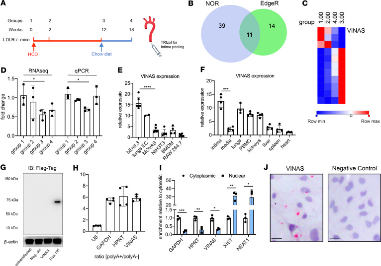
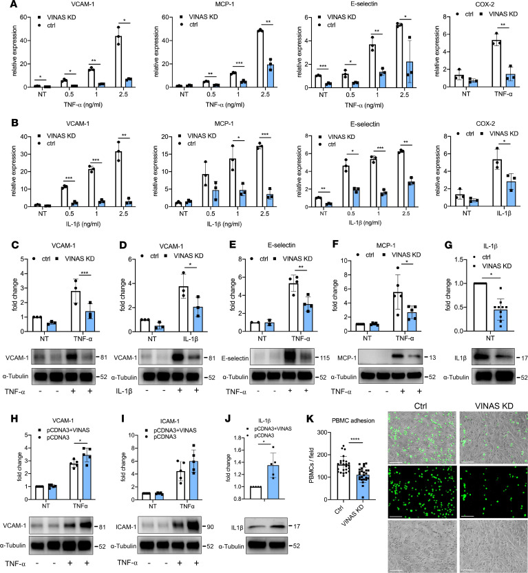
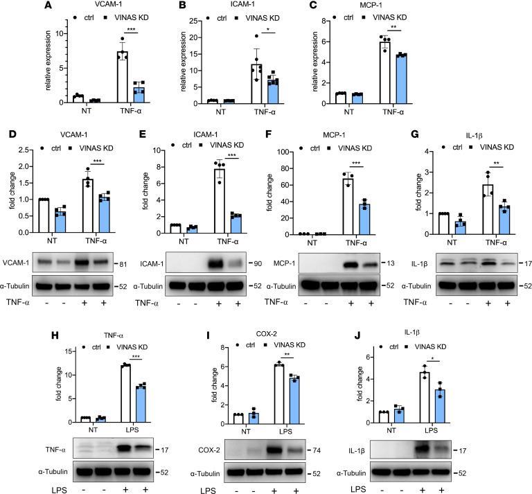
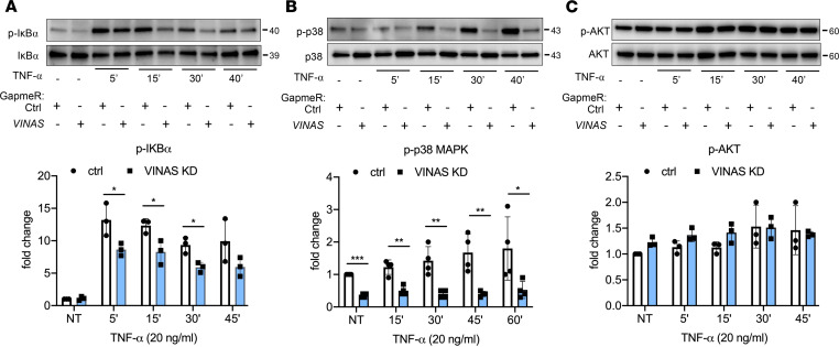
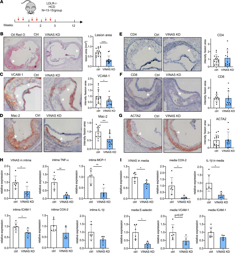
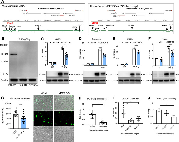
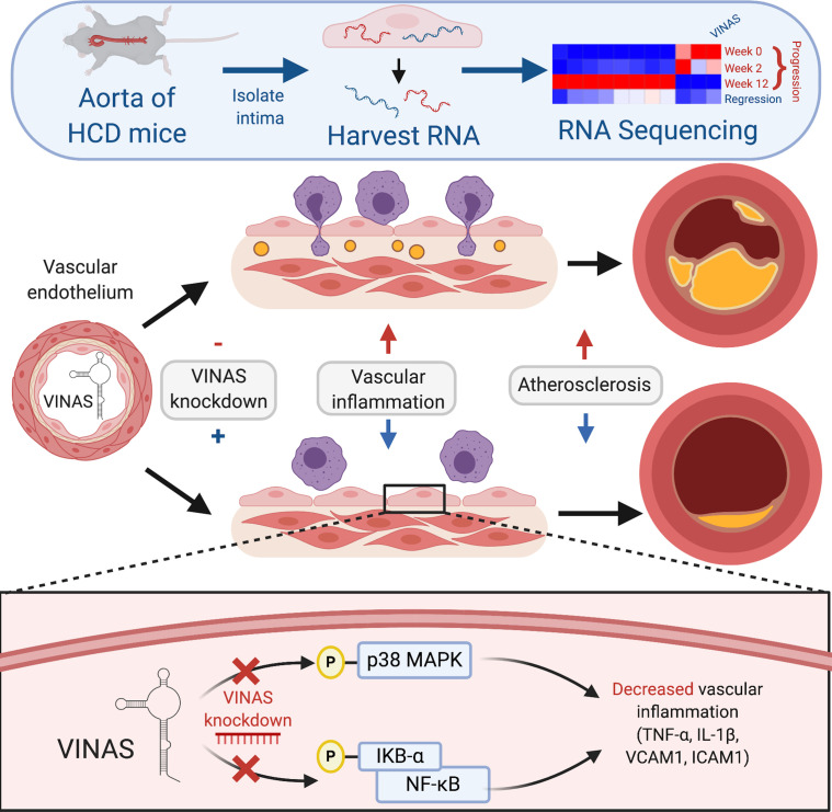
Long noncoding RNAs (lncRNAs) play important roles in regulating diverse cellular processes in the vessel wall, including atherosclerosis. RNA-Seq profiling of intimal lesions revealed a lncRNA, VINAS (Vascular INflammation and Atherosclerosis lncRNA Sequence), that is enriched in the aortic intima and regulates vascular inflammation. Aortic intimal expression of VINAS fell with atherosclerotic progression and rose with regression. VINAS knockdown reduced atherosclerotic lesion formation by 55% in LDL receptor-deficient (LDLR-/-) mice, independent of effects on circulating lipids, by decreasing inflammation in the vessel wall. Loss- and gain-of-function studies in vitro demonstrated that VINAS serves as a critical regulator of inflammation by modulating NF-κB and MAPK signaling pathways. VINAS knockdown decreased the expression of key inflammatory markers, such as MCP-1, TNF-α, IL-1β, and COX-2, in endothelial cells (ECs), vascular smooth muscle cells, and bone marrow-derived macrophages. Moreover, VINAS silencing decreased expression of leukocyte adhesion molecules VCAM-1, E-selectin, and ICAM-1 and reduced monocyte adhesion to ECs. DEP domain containing 4 (DEPDC4), an evolutionary conserved human ortholog of VINAS with approximately 74% homology, showed similar regulation in human and pig atherosclerotic specimens. DEPDC4 knockdown replicated antiinflammatory effects of VINAS in human ECs. These findings reveal a potentially novel lncRNA that regulates vascular inflammation, with broad implications for vascular diseases.

Keywords: Atherosclerosis; Noncoding RNAs; Vascular Biology; endothelial cells.


Conflict of interest statement

Conflict of interest: The authors have declared that no conflict of interest exists.

Figures

Figure 1
Figure 1. Identification of the lncRNA VINAS in lesional intima.
(A) RNA derived from aortic intima of LDLR–/– mice (n = 3; each sample represents RNA pooled from 2 mice) that were placed on a high-cholesterol diet (HCD) for 0 (group 1), 2 (group 2), 12 (group 3), and 18 weeks after 6 weeks of resumption of a normal chow diet (group 4). (B) Venn diagram displays significantly dysregulated lncRNAs in genome-wide RNA-Seq profiling using EdgeR and no-overlapping reads (NOR) showing intersecting hits (n = 11), uniquely identified in EdgeR (n = 14) or NOR (n = 39), (log2 fold change [1.5]; FDR < 0.05). (C) Heatmap for 11 lncRNAs that were dynamically regulated with progression and regression of atherosclerosis (n = 3). (D) RNA-Seq results for VINAS across groups 1–4 obtained by RNA-Seq analysis and verified by RT-qPCR (n = 3). (E) RT-qPCR expression analysis for VINAS in different cell types (n = 3). (F) VINAS expression in body organs and PBMCs of 24-week-old C57BL/6 mice (n = 4). (G) To test the coding potential, VINAS sequence was cloned upstream of 3xFlag-Tag cassette, transfected in HEK293T cells, and immunoblotted for Flag antibody. Positive control was provided with the kit (representative of 3 experiments). (H) RNA from mouse extracellular cells (ECs) was isolated for polyA+ and polyA enriched RNA and analyzed by real-time quantitative PCR (RT-qPCR) (n = 3). (I) RT-qPCR analysis for RNA derived from mouse ECs separated into cytoplasmic and nuclear fractions and normalized to the cytoplasmic fraction (n = 3). (J) RNA in situ hybridization for negative control and VINAS probes on paraformaldehyde-fixed mouse ECs. Scale bar: 5 μm. Data represent the mean ± SD. Statistical differences were calculated using unpaired 2-tailed Student’s t test except for multiple comparisons (E and F) in which 1-way ANOVA with Bonferroni’s correction was used. *P < 0.05, **P < 0.01, ***P < 0.001.
Figure 2
Figure 2. VINAS regulates inflammatory markers in endothelial cells.
VINAS knockdown decreases the mRNA levels of VCAM-1, E-selectin, MCP-1, and COX2 in mouse ECs activated with TNF-α (A) and IL-1β (B); n = 3. VINAS silencing decreases the protein expression of VCAM-1 (C and D, n = 3), E-selectin (E, n = 4), MCP-1 (F, n = 5), and IL-1β (G, n = 10) in basal conditions or after activation with 20 ng/mL TNF- or IL-1β. VINAS overexpression increases the protein expression of VCAM-1 (H), ICAM-1 (I), and IL-1β (J) in mouse ECs not treated or activated with 20 ng/mL TNF-α (n = 5). (K) VINAS knockdown decreases the PBMCs’ adhesion to mouse ECs activated with TNF-α for 4 hours (5 ng/mL, representative of 3 experiments). Scale bar: 50 μm. Data represent the mean ± SD. Statistical differences were calculated using unpaired 2-tailed Student’s t test. *P < 0.05, **P < 0.01, ***P < 0.001, ****P < 0.0001.
Figure 3
Figure 3. VINAS knockdown decreases inflammation in SMCs and BMDMs.
VINAS knockdown decreases mRNA levels of VCAM-1 (A, n = 4), ICAM-1 (B, n = 6), and MCP-1 (C, n = 4) in MOVAS smooth muscle cells (SMCs) stimulated with 5 ng/mL TNF-α. VINAS knockdown decreases protein expression of VCAM-1 (D, n = 4), ICAM-1 (E, n = 4), MCP-1 (F, n = 3), and IL-1β (G, n = 4) in MOVAS smooth muscle cells stimulated with 20 ng/mL TNF-α. VINAS knockdown decreases the protein expression of TNF-α (H), COX-2 (I), and IL-1β (J) in bone marrow–derived macrophages (BMDMs) stimulated with 50 ng/mL LPS (n = 3). Data represent the mean ± SD. Statistical differences were calculated using unpaired 2-tailed Student’s t test. *P < 0.05, **P < 0.01, ***P < 0.001.
Figure 4
Figure 4. VINAS knockdown regulates NF-κB and p38 MAPK signaling pathways.
Mouse ECs were transfected with VINAS gapmeRs and activated with TNF-α (20 ng/mL) for 5, 15, 30, 45, and 60 minutes. Phosphorylation of IκBα (A, n = 3), p38 MAPK (B, n = 4), and AKT (C, n = 3) was assessed by Western blot. Data represent the mean ± SD. Statistical differences were calculated using 1-way ANOVA with Bonferroni’s correction. *P < 0.05, **P < 0.01, ***P < 0.001.
Figure 5
Figure 5. In vivo knockdown of VINAS inhibits atherosclerotic lesion formation by decreasing vascular inflammation.
(A) LDLR/ mice were i.v. injected with vehicle control gapmeR (n = 15) or VINAS gapmeR (n = 13) twice per week (10 mg/kg/mouse/injection) and placed on an HCD for 12 weeks. Representative images and quantification for Oil Red O (scale bar: 400 μm) (B), VCAM-1 (C), Mac-2 (D), CD4+ (E), CD8+ (F), and ACTA2 (G) staining (arrowhead) of the aortic sinus of LDLR–/– HCD mice treated with control (n = 15) or MAARS (n = 13) gapmeRs for 12 weeks. Scale bar: 100 μm. VINAS silencing efficiency and expression of inflammatory markers was assessed by RT-qPCR in the intima (H) and media (I) fractions of the aortic arch from control gapmeR (n = 6) and VINAS gapmeR groups (n = 5). Data represent the mean ± SD. Statistical differences were calculated using unpaired 2-tailed Student’s t test. *P < 0.05, **P < 0.01, ***P < 0.001, ****P < 0.0001.
Figure 6
Figure 6. DEPDC4 is a human ortholog of VINAS.
(A) Illustration of the genomic locations of VINAS and DEPDC4 in the mouse and human chromosomes 10 and 12, respectively. (B) DEPDC4 does not encode for a protein or peptide. To test the coding potential, DEPDC4 sequence was cloned upstream of the 3xFlag-Tag cassette, transfected in HEK293T cells, and immunoblotted for Flag antibody; positive control was provided with the kit (n = 3 experiments). DEPDC4 silencing decreases the protein expression of VCAM-1 (C, n = 7), E-selectin (D, n = 5), and ICAM-1 (E, n = 3) COX-2 (F, n = 6) in HUVECs activated with 20 ng/mL TNF-α. (G) DEPDC4 knockdown decreases THP-1 monocyte adhesion to HUVEC monolayers activated with TNF-α for 4 hours (5 ng/mL, representative images and quantification of adhered monocytes). (H) RT-qPCR of DEPDC4 in human carotid arteries with stable (n = 6) or unstable (n = 7) atherosclerotic plaques. Scale bar: 50 μm. (I) Expression of DEPDC4 from RNA-Seq analyses of lesions with increasing severity of coronary atherosclerosis in Yorkshire pigs fed an HCD for 60 weeks (n = 4/group). (J) RT-qPCR of VINAS expression in aortic intima of LDLR–/– mice at 0, 2, and 12 weeks of an HCD (n = 3/group). Data represent the mean ± SD. Statistical differences were calculated using unpaired 2-tailed Student’s t test except for multiple comparisons (I and J) in which 1-way ANOVA with Bonferroni’s correction was used. *P < 0.05, **P < 0.01, ***P < 0.001, ****P < 0.0001.
Figure 7
Figure 7. Summary of the role of lncRNA VINAS in vascular inflammation.
RNA-Seq profiling of intimal lesions revealed VINAS lncRNA that is enriched in the aortic intima, decreased with atherosclerotic progression, and increased with regression. VINAS knockdown decreased the expression of key inflammatory markers, NF-κB and MAPK signaling pathways, cell adhesion molecules, and the monocytes adhesion to ECs. In vivo VINAS knockdown reduced atherosclerotic lesion formation in LDLR–/– mice by decreasing vascular inflammation.

References

    1. Libby P. Inflammation in atherosclerosis. Arterioscler Thromb Vasc Biol. 2012;32(9):2045–2051. doi: 10.1161/ATVBAHA.108.179705. - DOI - PMC - PubMed
    1. Wolf D, Ley K. Immunity and inflammation in atherosclerosis. Circ Res. 2019;124(2):315–327. doi: 10.1161/CIRCRESAHA.118.313591. - DOI - PMC - PubMed
    1. Virchow R. Cellular pathology. As based upon physiological and pathological histology. Lecture XVI--Atheromatous affection of arteries. 1858. Nutr Rev. 1989;47(1):23–25. - PubMed
    1. Ridker PM, et al. Antiinflammatory therapy with canakinumab for atherosclerotic disease. N Engl J Med. 2017;377(12):1119–1131. doi: 10.1056/NEJMoa1707914. - DOI - PubMed
    1. Nahrendorf M. Myeloid cell contributions to cardiovascular health and disease. Nat Med. 2018;24(6):711–720. doi: 10.1038/s41591-018-0064-0. - DOI - PMC - PubMed

Publication types

MeSH terms