. 2020 Nov 5;107(5):882-894.
doi: 10.1016/j.ajhg.2020.09.006. Epub 2020 Oct 5.

Plasma DNA Profile Associated with DNASE1L3 Gene Mutations: Clinical Observations, Relationships to Nuclease Substrate Preference, and In Vivo Correction

Affiliations

Plasma DNA Profile Associated with DNASE1L3 Gene Mutations: Clinical Observations, Relationships to Nuclease Substrate Preference, and In Vivo Correction

Rebecca W Y Chan et al. Am J Hum Genet. .

Erratum in

Abstract

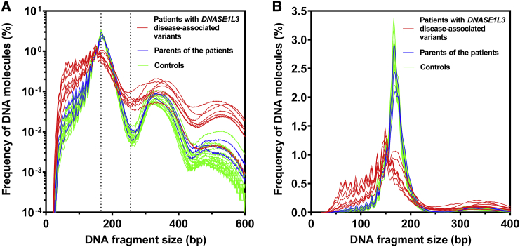
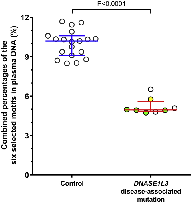
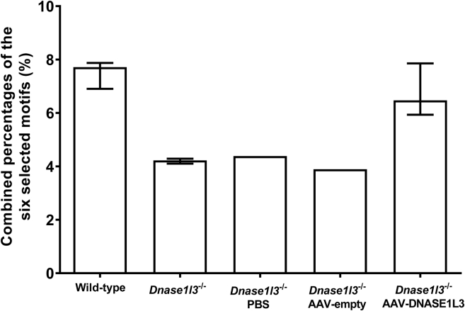
Plasma DNA fragmentomics is an emerging area in cell-free DNA diagnostics and research. In murine models, it has been shown that the extracellular DNase, DNASE1L3, plays a role in the fragmentation of plasma DNA. In humans, DNASE1L3 deficiency causes familial monogenic systemic lupus erythematosus with childhood onset and anti-dsDNA reactivity. In this study, we found that human patients with DNASE1L3 disease-associated gene variations showed aberrations in size and a reduction of a "CC" end motif of plasma DNA. Furthermore, we demonstrated that DNA from DNASE1L3-digested cell nuclei showed a median length of 153 bp with CC motif frequencies resembling plasma DNA from healthy individuals. Adeno-associated virus-based transduction of Dnase1l3 into Dnase1l3-deficient mice restored the end motif profiles to those seen in the plasma DNA of wild-type mice. Our findings demonstrate that DNASE1L3 is an important player in the fragmentation of plasma DNA, which appears to act in a cell-extrinsic manner to regulate plasma DNA size and motif frequency.

Keywords: autoimmune disease; biomarkers; cfDNA; circulating DNA; liquid biopsy; systemic lupus erythematosus.


Conflict of interest statement

R.W.Y.C., M.N., P.J., A.S.H.C., W.P., D.S.C.H., K.C.A.C., R.W.K.C., and Y.M.D.L. are co-inventors on multiple patents in cell-free DNA-based diagnostics. Y.M.D.L. is a scientific co-founder, shareholder, and scientific advisor of Grail. R.W.K.C. and K.C.A.C. are shareholders of Grail. Y.M.D.L., R.W.K.C., and K.C.A.C. are co-founders and shareholders of DRA Limited and the Take2 Group of companies. Y.M.D.L. is an advisor to Decheng Capital. R.W.K.C. is an advisor to Illumina. Y.M.D.L., R.W.K.C., and K.C.A.C. receive royalties from Illumina, Sequenom, DRA, Take2, and Grail.

Figures

Figure 1
Figure 1
Conceptual Diagram Showing the Design and Experimental Approaches of the Study In part I, we aimed to study the fragmentomic characteristics of plasma DNA from human individuals with or without disease-associated variations of DNASE1L3. Plasma DNA was isolated from the blood of subjects with DNASE1L3 disease-associated variants, their parents, and control subjects for analysis and comparison. In part II, we aimed to study the fragmentation preference of DNASE1L3 and DNASE1 by in vitro digestion of DNA from a human cell line. DNA from cell nuclei and naked DNA, representing DNA with or without nucleosomal structural proteins, respectively, was subjected to enzymatic digestion by DNASE1L3 or DNASE1 prior to sequence analysis. In part III, we used a mouse model to investigate whether the fragmentomic profile of DNA in plasma of Dnase1l3-deficient mice could be corrected in vivo via DNASE1L3-expressing adeno-associated viral (AAV) transfection. Dnase1l3-deficient mice injected with empty AAV particles or PBS were used as negative controls. The DNA libraries were constructed and sequenced. The fragmentomic profiling involved the analyses of the size distribution and the 4-mer end motif frequencies of DNA fragments. The red cross denotes the inefficiency of DNASE1 in digestion of cell nuclei (refer to Figure 7).
Figure 2
Figure 2
Size Distributions of Plasma DNA Molecules of Case Subjects, Their Parents, and Control Subjects Subjects with DNASE1L3 disease-associated variants (red, n = 8), their parents (blue, n = 3), and control subjects (green, n = 19). The size distribution is plotted in the range (A) from 0 to 600 bp on a logarithmic scale and (B) from 0 to 400 bp on a linear scale. Each line represents one case. As one subject provided four blood collections, a total of eight plasma samples from the affected group were analyzed.
Figure 3
Figure 3
Fragmentation Index of DNA in Plasma of Control Subjects and Case Subjects with DNASE1L3 Disease-Associated Variants The fragmentation index was calculated by dividing the sequenced read count of the DNA molecules at 166 bp by that of the DNA molecules at 250 bp and plotted on a logarithmic scale. Each circle represents one case. As one subject provided four blood collections, a total of eight plasma samples from the affected group were analyzed. Multiple samples obtained from the same subject were indicated by green solid circles. One of the subjects (A3) with the missense mutation c.563G>C (p.Gln188Arg) was indicated by a yellow solid circle. The middle line and error bars indicate median ± interquartile range.
Figure 4
Figure 4
End Motif Aberrations of Plasma DNA in Subjects with DNASE1L3 Disease-Associated Variants Heatmap of the frequency of the top 25 ranked 4-mer end motifs in plasma DNA among the different groups. Samples V1 to V4 were taken on four separate occasions, from one patient who was homozygous for the c.290_291delCA (p.Thr97Ilefs2) frameshift mutation. Samples A3, H2, and H4 were taken from subjects who were also homozygous for the c.290_291delCA (p.Thr97Ilefs2) frameshift mutation. Sample A3 was taken from a subject who was homozygous for c.563G>C (p.Gln188Arg), and V5 was taken from a subject who was heterozygous for c.290_291delCA (p.Thr97Ilefs2) and a deletion in exon 5 of DNASE1L3. Sample H1 was taken from a parent who was heterozygous for c.563G>C (p.Gln188Arg), while samples H2 and H4 were taken from parents who were heterozygous for c.290_291delCA (p.Thr97Ilefs2) of DNASE1L3. SLE, systemic lupus erythematosus; JIA, juvenile idiopathic arthritis; transplant, subjects with a history of kidney transplantation; parent, healthy parents of subjects with DNASE1L3 disease-associated variants.
Figure 5
Figure 5
Combined Percentages of the Six Selected Motifs in Plasma DNA of Control Subjects and Case Subjects with DNASE1L3 Disease-Associated Variants The selected motifs CCCA, CCAG, CCTG, CCAA, CCCT, and CCAT were the top six 4-mer motifs ranked with the highest frequencies in healthy control subjects which showed a statistically significant reduction in case subjects with DNASE1L3 disease-associated variants. Each circle represents one case. As one subject provided blood samples on four different occasions, a total of eight plasma samples from the patient group were analyzed. Multiple samples obtained from the same subject were indicated by green solid circles. One of the subjects (A3) with the missense mutation c.563G>C (p.Gln188Arg) was indicated by a yellow solid circle. The middle line and error bars indicate median ± interquartile range.
Figure 6
Figure 6
Size Distributions of Nuclei DNA and Naked DNA after Digestion by Nucleases In Vitro The size distributions are in the range from 0 to 400 bp plotted on a linear scale.
Figure 7
Figure 7
Relationships between the Motif Rankings of In Vitro Digested DNA Substrates Correlations between (A) the motif rankings of cell nuclei after digestion with DNASE1L3 and that of cell nuclei incubated with DNASE1 and (B) the motif rankings of naked DNA digestion by DNASE1L3 and that of naked DNA digestion by DNASE1. Each circle represents a 4-mer motif. Motifs preferentially created by the DNASE1L3 digestion of cell nuclei (A) or naked DNA (B) were indicated by open red circles. Motifs preferentially created by the DNASE1 digestion of cell nuclei (A) or naked DNA (B) were indicated by open blue circles. Two common motifs preferentially created by DNASE1 digestion of both cell nuclei and naked DNA were indicated by solid blue circles.
Figure 8
Figure 8
Heatmap Showing the Effect of DNASE1L3 Reconstitution on the 4-mer Motifs of Plasma DNA in Dnase1l3-Deficient Mice The top 25 4-mer motifs with the highest frequencies in the wild-type mice were compared. Publicly available data of 9 wild-type and 13 Dnase1l3-deficient mice were used as references.
Figure 9
Figure 9
Combined Percentages of the Six Selected Motifs in Plasma DNA of Mice with or without DNASE1L3 Reconstitution The selected motifs CCCA, CCTG, CCAG, CCAA, CCAT, and CCTC were the top six 4-mer motifs ranked with the highest frequencies in wild-type mice and showed a statistically significant reduction in Dnase1l3-deficient mice. Publicly available data from 9 wild-type and 13 Dnase1l3-deficient mice were used as references. The bar and errors indicate median ± interquartile range.

References

    1. Chiu R.W.K., Chan K.C.A., Gao Y., Lau V.Y.M., Zheng W., Leung T.Y., Foo C.H.F., Xie B., Tsui N.B.Y., Lun F.M.F. Noninvasive prenatal diagnosis of fetal chromosomal aneuploidy by massively parallel genomic sequencing of DNA in maternal plasma. Proc. Natl. Acad. Sci. USA. 2008;105:20458–20463. - PMC - PubMed
    1. Chiu R.W.K., Akolekar R., Zheng Y.W.L., Leung T.Y., Sun H., Chan K.C.A., Lun F.M.F., Go A.T.J.I., Lau E.T., To W.W.K. Non-invasive prenatal assessment of trisomy 21 by multiplexed maternal plasma DNA sequencing: large scale validity study. BMJ. 2011;342:c7401. - PMC - PubMed
    1. Cohen J.D., Li L., Wang Y., Thoburn C., Afsari B., Danilova L., Douville C., Javed A.A., Wong F., Mattox A. Detection and localization of surgically resectable cancers with a multi-analyte blood test. Science. 2018;359:926–930. - PMC - PubMed
    1. Chan K.C.A., Woo J.K.S., King A., Zee B.C.Y., Lam W.K.J., Chan S.L., Chu S.W.I., Mak C., Tse I.O.L., Leung S.Y.M. Analysis of plasma Epstein–Barr Virus DNA to screen for nasopharyngeal cancer. N. Engl. J. Med. 2017;377:513–522. - PubMed
    1. Chan K.C.A., Zhang J., Hui A.B.Y., Wong N., Lau T.K., Leung T.N., Lo K.W., Huang D.W.S., Lo Y.M.D. Size distributions of maternal and fetal DNA in maternal plasma. Clin. Chem. 2004;50:88–92. - PubMed

Publication types

MeSH terms