. 2020 Nov 5;80(3):423-436.e9.
doi: 10.1016/j.molcel.2020.09.017. Epub 2020 Oct 5.

Ubiquitin Phosphorylation at Thr12 Modulates the DNA Damage Response

Affiliations

Ubiquitin Phosphorylation at Thr12 Modulates the DNA Damage Response

Franziska Walser et al. Mol Cell. .

Abstract

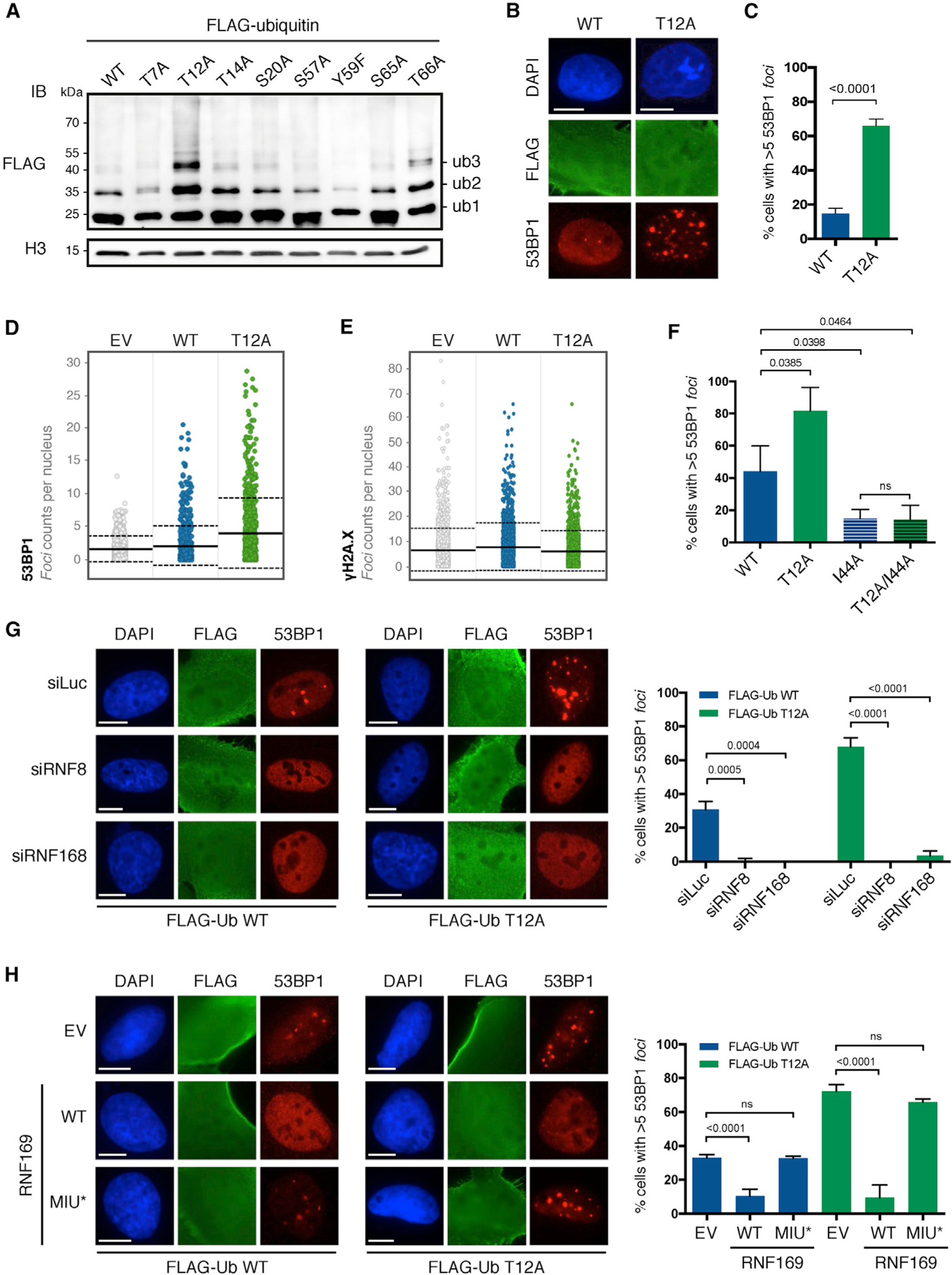
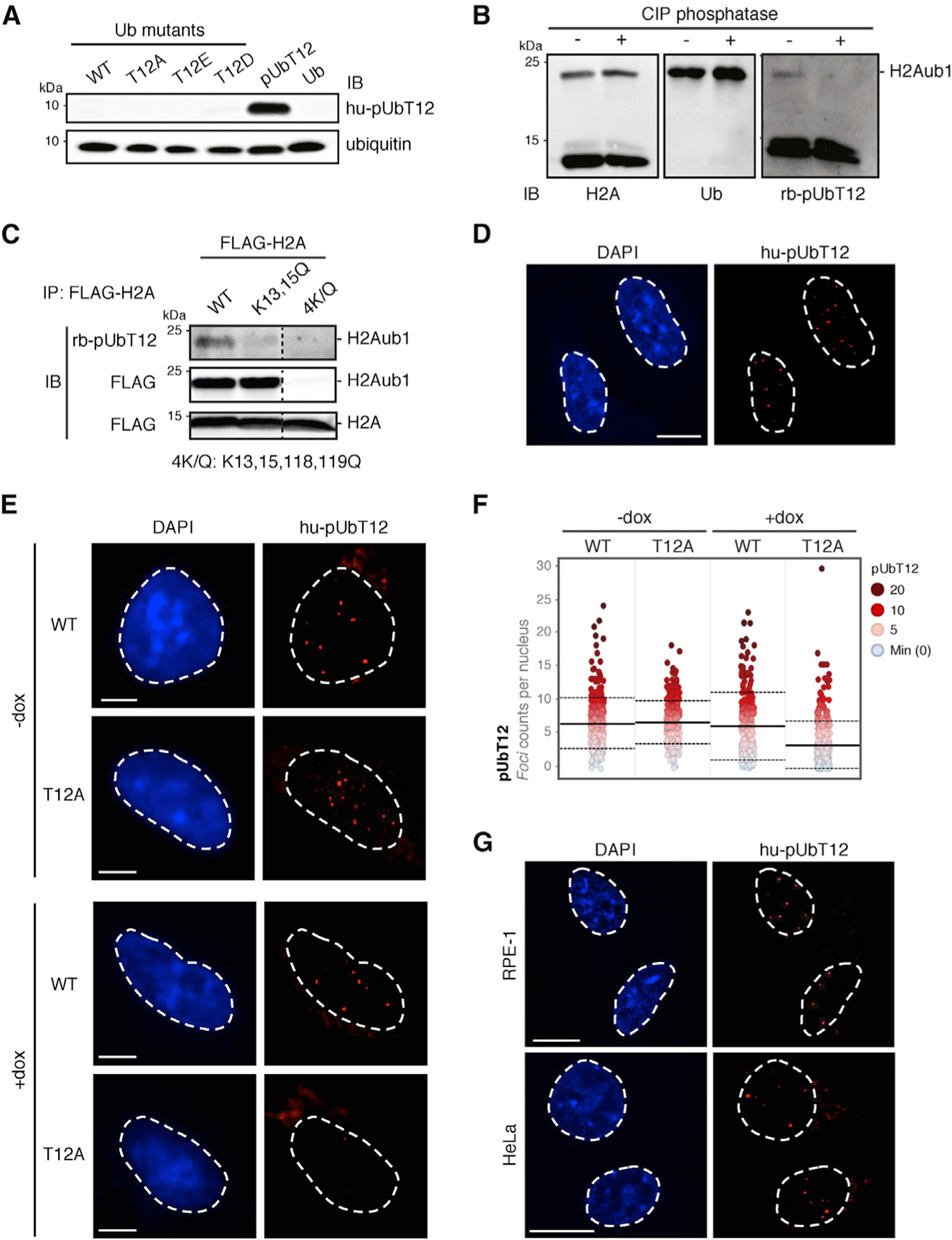
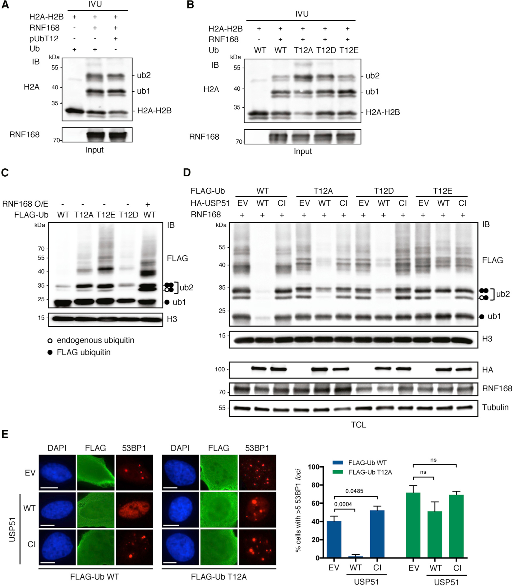
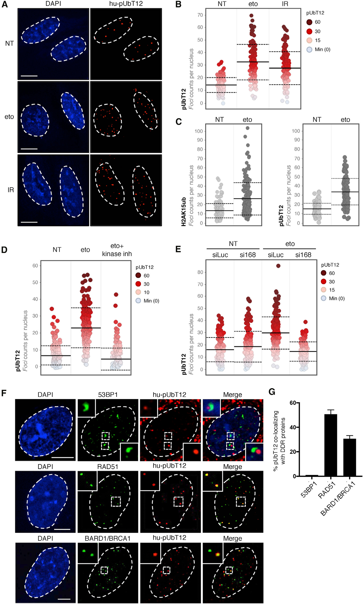
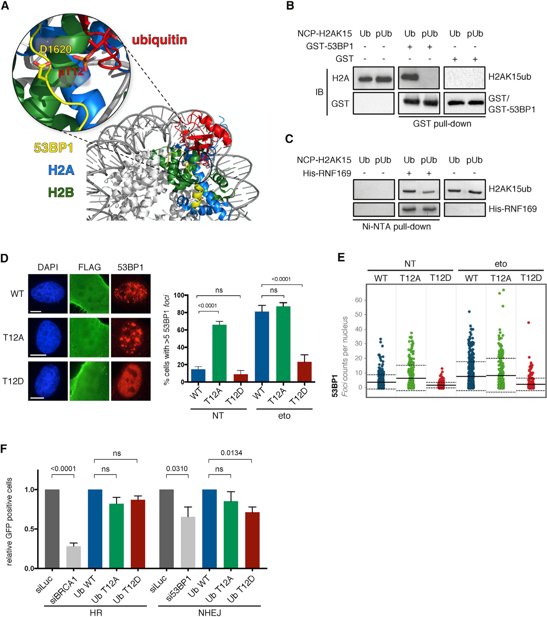
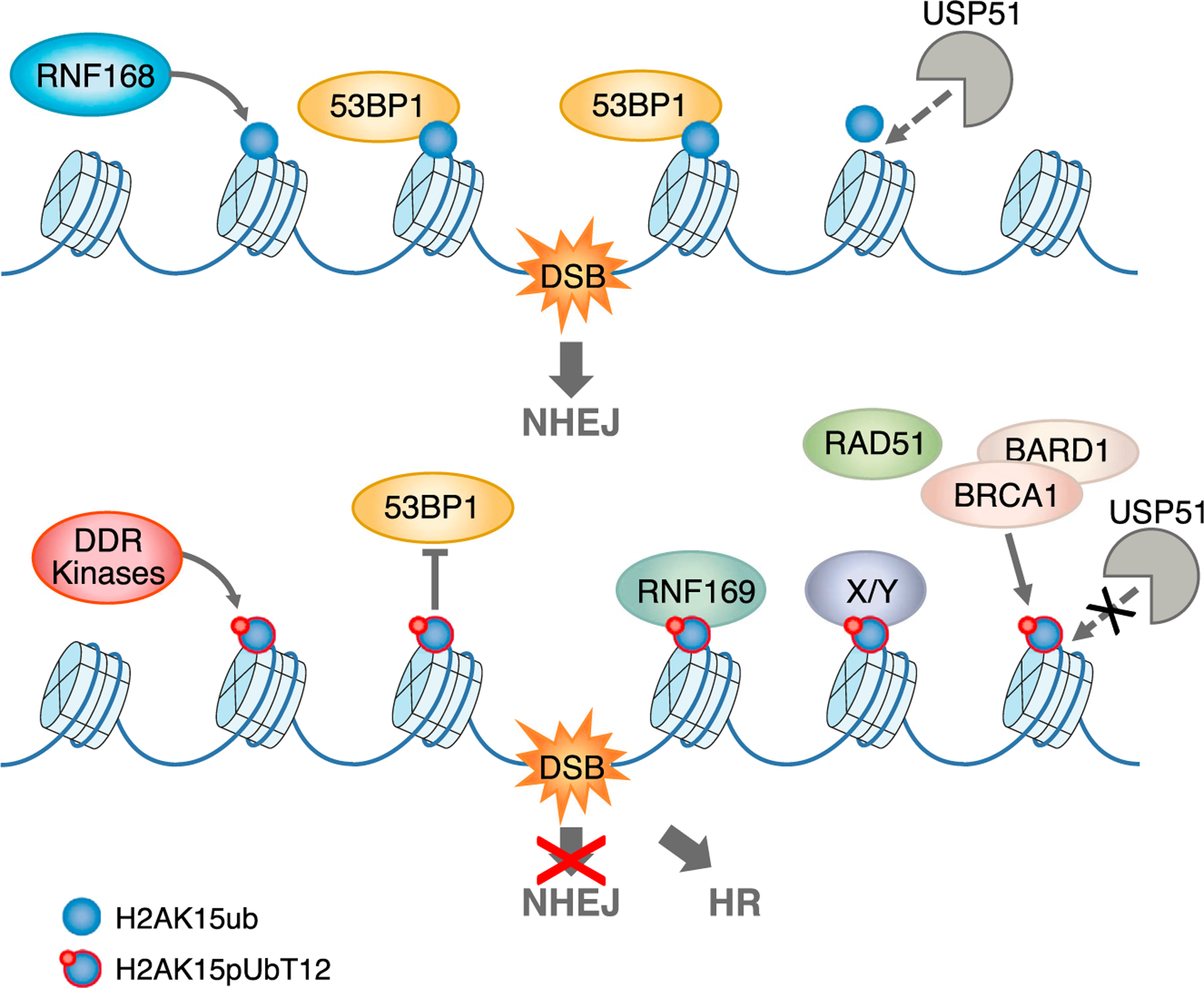
The ubiquitin system regulates the DNA damage response (DDR) by modifying histone H2A at Lys15 (H2AK15ub) and triggering downstream signaling events. Here, we find that phosphorylation of ubiquitin at Thr12 (pUbT12) controls the DDR by inhibiting the function of 53BP1, a key factor for DNA double-strand break repair by non-homologous end joining (NHEJ). Detectable as a chromatin modification on H2AK15ub, pUbT12 accumulates in nuclear foci and is increased upon DNA damage. Mutating Thr12 prevents the removal of ubiquitin from H2AK15ub by USP51 deubiquitinating enzyme, leading to a pronounced accumulation of ubiquitinated chromatin. Chromatin modified by pUbT12 is inaccessible to 53BP1 but permissive to the homologous recombination (HR) proteins RNF169, RAD51, and the BRCA1/BARD1 complex. Phosphorylation of ubiquitin at Thr12 in the chromatin context is a new histone mark, H2AK15pUbT12, that regulates the DDR by hampering the activity of 53BP1 at damaged chromosomes.

Keywords: 53BP1; BRCA1/BARD1; DDR; DNA damage response; DNA repair; H2AK15pUbT12; RAD51; RNF168; RNF169; RNF8; USP51; chromatin ubiquitination; genome stability; histone mark H2AK15ub; pUbT12; phospho-ubiquitin Thr12; ubiquitin phosphorylation.


Conflict of interest statement

Declaration of Interests H.O. is a shareholder of UbiQ B.V.

Figures

Figure 1.
Figure 1.. UbT12 Regulates 53BP1 Foci Formation, which Depends on RNF8 and RNF168 and Is Counteracted by RNF169
(A) Chromatin ubiquitination in HEK293T cells upon 24-h overexpression of the indicated FLAG-ubiquitin wild-type (WT) and point mutants. After acidic extraction, chromatin fractions were analyzed by FLAG and H3 immunoblot (IB). Ubiquitinated forms of histones are indicated (ub1, ub2, and ub3). (B and C) 53BP1 foci formation in untreated U2OS cells after 24-h transfection of FLAG-ubiquitin WT or T12A and immunostained for FLAG, 53BP1, and DAPI. Representative images (B) and quantification of cells with more than five 53BP1 foci (C) in FLAG-positive cells are shown. Scale bars, 10 μm (B). At least 50 cells per condition were counted in each replicate, and data are represented as mean + SD (n = 3). (D and E) Quantitative image-based cytometry (QIBC) shows the distribution of 53BP1 foci (D) and γH2A.X foci (E) in FLAG-positive cells transfected and stained as in (B). EV, empty vector. For each condition, images containing at least 1,000 cells per experiment were acquired (n = 3). Mean (solid line) and SD from the mean (dashed lines) are indicated. (F–H) Quantification of cells with more than five 53BP1 foci in FLAG-positive U2OS cells transfected for 24 h with indicated FLAG-ubiquitin mutants (n = 3) (F) upon 72-h knockdown of RNF8 and RNF168 (siRNF8 and siRNF168; n = 3) (G) and upon 48-h overexpression of RNF169 WT or MIU-defective mutant (MIU*; n = 3) (H). Cells were immunostained for FLAG, 53BP1, and DAPI. At least 50 cells per condition were counted, and data are represented as mean + SD. Scale bars, 10 μm. See also Figure S1.
Figure 2.
Figure 2.. UbT12 Is Phosphorylated in Chromatin Extracts at H2AK15ub and Accumulates in Nuclear Foci
(A) Validation of pUbT12-specific antibody immunoglobulin G1 (IgG1) (clone 2.B5, referred as hupUbT12) by IB on chemically synthesized full-length pUbT12 and recombinant ubiquitin (Ub) mutants expressed in E. coli, as indicated. (B) pUbT12 was detected in chromatin fraction of HEK293T cells by pUbT12 IB (H2Aub1; rb-pUbT12). H2A and ubiquitin IB are used as reference. Fractions were treated with calf intestinal phosphatase (CIP) to assess the specificity of the signal. (C) Chromatin fractions of HEK293T cells transfected with FLAG-H2A WT and the indicated mutants were immunoprecipitated with FLAG antibody. pUbT12 and FLAG signals were detected by IB, and irrelevant lanes were digitally removed from the blot image (dashed line). Mono-ubiquitinated (H2Aub1) and unmodified H2A are indicated. (D) Confocal analysis of nuclear pUbT12 foci in untreated U2OS cells stained with hu-pUbT12 and DAPI. Scale bar, 10 μm. (E and F) pUbT12 foci in U2OS WT and T12A ubiquitin replacement cells, after 4 days of doxycycline (dox) induction, were analyzed by hu-pUbT12 and DAPI staining. Representative images (E) and quantification of nuclear pUbT12 foci (F) are shown. Scale bars, 5 μm (E). Each dot (F) represents a single cell color-coded according to pUbT12 foci number. For each condition, images containing at least 150 cells per experiment were acquired (n = 3). Mean (solid line) and SD from the mean (dashed lines) are indicated. (G) Confocal analysis of nuclear pUbT12 foci in untreated RPE-1 and HeLa cells stained with hupUbT12 and DAPI. Scale bars, 10 μm. See also Figure S2.
Figure 3.
Figure 3.. pUbT12 Can Be Conjugated to Nucleosomes but Cannot be Cleaved by the Deubiquitinating Enzyme USP51
(A and B) In vitro ubiquitination assay (IVU) using purified E1, E2 (UbcH5c), E3 (GST-RNF168), and H2A-H2B dimer, together with ubiquitin (Ub), chemically synthetized pUbT12 (A) or bacterially expressed ubiquitin mutants (B), as indicated. Ubiquitinated H2A-H2B was detected by IB using anti-H2A antibody; unmodified, mono- and di-ubiquitinated forms of H2A-H2B dimer are indicated (H2A-H2B, ub1, and ub2). (C) Chromatin ubiquitination in HEK293T cells upon 24-h overexpression of the indicated FLAG-ubiquitin forms together with RNF168 (RNF168 O/E; +) or the empty vector (−). After acidic extraction, chromatin fractions were analyzed by FLAG and H3 IB. Ubiquitinated forms of histones, modified by endogenous (white circle) or exogenous (FLAG tagged; black circle) are indicated (ub1 and ub2). (D) IB of HEK293T chromatin extracts upon 48-h overexpression of RNF168 and the indicated FLAG-ubiquitin forms, together with USP51 WT or the catalytic inactive (CI) mutant. TCL, total cell lysate; EV, empty vector. (E) 53BP1 foci formation in untreated U2OS cells upon 24-h overexpression of FLAG-ubiquitin WT or T12A and USP51 WT or CI mutant, stained for FLAG, 53BP1, and DAPI. Representative images (left) and quantification of cells with more than five 53BP1 foci (right) in FLAG-positive cells are shown. Scale bars, 10 μm. At least 50 cells per condition were counted in each replicate, and data are represented as mean + SD (n = 3). See also Figure S3.
Figure 4.
Figure 4.. pUbT12 Is Induced by DNA Damage and Depends on RNF168
(A and B) Confocal analysis (A) and quantification (B) showing the distribution of nuclear pUbT12 foci in U2OS cells treated for 1 h with etoposide (eto; 5 μM), irradiated (IR; 1 Gy), or left untreated (NT). Each dot (B) represents a single cell color-coded according to pUbT12 foci number. Scale bars, 10 μm. For each condition, images containing at least 150 cells per experiment were acquired (n = 3). Mean (solid line) and SD from the mean (dashed lines) are indicated. (C) Quantification of nuclear H2AK15ub and pUbT12 stained foci in U2OS cells treated for 1 h with 5 μM etoposide or left untreated. Each dot represents a single cell, and images containing at least 150 cells per condition were acquired. Mean (solid line) and SD from the mean (dashed lines) are indicated. (D) Quantification of nuclear pUbT12 foci in U2OS cells treated for 1 h with 5 μM etoposide, left untreated, or pretreated with kinase inhibitors (ATM inhibitor KU-55933, 10 μM; ATR inhibitor VE-821, 10 μM; and DNA-PK inhibitor KU-57788, 2 μM) for 2 h before etoposide treatment. Each dot represents a single cell color-coded according to pUbT12 foci number. For each condition, images containing at least 150 cells per experiment were acquired (n = 3). Mean (solid line) and standard deviation from the mean (dashed lines) are indicated. (E) Quantification showing the distribution of nuclear pUbT12 foci in U2OS cells treated for 1 h with 5 μM etoposide after 96-h depletion of RNF168 (si168). siLuc served as a control. Each dot represents a single cell color-coded according to pUbT12 foci number. For each condition, images containing at least 150 cells per experiment were acquired (n = 3). Mean (solid line) and SD from the mean (dashed lines) are indicated. (F and G) Confocal analysis of co-localization between pUbT12-positive foci and 53BP1, RAD51, or BARD1 in U2OS cells treated for 1 h with etoposide (5 μM). Representative images (F) and quantification of colocalizing signals (G) are shown. Scale bars, 5 μm. At least 250 pUbT12-positive foci per condition were counted in each replicate (n = 3). See also Figure S4.
Figure 5.
Figure 5.. pUbT12 Limits Recruitment and Function of 53BP1 to Damaged Chromatin
(A) Structural prediction of the interference of ubiquitin phosphorylation at Thr12 in the binding of 53BP1 with the nucleosome, using a cryoelectron microscopy model (NCP-H2AK15ub-53BP1). (B) H2A and GST IB of GST pull-down assays using GST or GST-53BP1 with nucleosomes modified on H2AK15 by ubiquitin (Ub) or pUbT12 (pUb). (C) Coomassie blue staining of Ni-NTA pull-down assays with NCP-H2AK15ub using Ub or pUbT12 and His-RNF169. (D) 53BP1 foci formation in untreated (NT) and etoposide (eto) treated (1 h, 5 μM) U2OS cells upon 48-h FLAG-ubiquitin overexpression and 18-h ubiquitin knockdown immunostained for FLAG, 53BP1, and DAPI. Representative images (left) and quantification of cells with more than five 53BP1 foci (right) in FLAG-positive cells are shown (n = 3). At least 50 cells per condition were counted in each replicate, and data are represented as mean + SD (n = 3). Scale bars, 10 μm. (E) Nuclear 53BP1 foci analyzed by high-content microscopy in cells as in (D). For each condition, images containing at least 1,000 cells per experiment were acquired (n = 3). Mean (solid line) and SD from the mean (dashed lines) are indicated. (F) U2OS reporter cells, stably integrated with DR-GFP or EJ5-GFP to measure HR and NHEJ, respectively, were transfected with I-SceI and transduced with lentivirus (MOI = 1) for the indicated ubiquitin forms. For knockdown of BRCA1 (siBRCA1), 53BP1 (si53BP1), and control (siLuc), cells were transfected twice (72 h and 24 h before I-SceI transfection) with small interfering RNA (siRNA). After 72 h of I-SceI overexpression, GFP-positive cells were analyzed by FACS, and relative changes to ubiquitin WT or siLuc were calculated (n = 3). Data are represented as mean + SEM. See also Figures S5 and S6.
Figure 6.
Figure 6.. Model Depicting the Regulation of DDR Events on Chromatin Mediated by pUbT12
Top: DNA double-strand break (DSB) induces RNF168-dependent ubiquitination of histone H2A at Lys15 (H2AK15ub, blue sphere), which is directly bound by 53BP1, leading to DNA repair via non-homologous end joining (NHEJ). The deubiquitinating enzyme USP51 removes this mark, which attenuates the signaling, once the damage is repaired. Bottom: ubiquitin phosphorylation of H2AK15ub at Thr12, which depends on the upstream DDR kinases, generates the new epigenetic mark H2AK15pUbT12 (blue sphere with red circle), which impedes the recruitment of 53BP1 to damaged chromatin, blocking DNA repair via NHEJ. Importantly, H2AK15pUbT12 is permissive to the binding of RNF169 and the recruitment of RAD51 and the BRCA1/BARD1 complex, allowing DNA repair via homologous recombination (HR). Alteration of H2AK15ub by pUbT12 prevents the clearance of ubiquitin from chromatin by USP51, leading to sustained chromatin (phospho)ubiquitination. Proteins X/Y represent hypothetic factors that specifically recognize the H2AK15pUbT12 mark.

Similar articles

Cited by

References

    1. Ai H, Guo Y, Sun D, Liu S, Qi Y, Guo J, Qu Q, Gong Q, Zhao S, Li J, and Liu L (2019). Examination of the deubiquitylation site selectivity of USP51 by using chemically synthesized ubiquitylated histones. ChemBioChem 20, 221–229. - PubMed
    1. An L, Dong C, Li J, Chen J, Yuan J, Huang J, Chan KM, Yu CH, and Huen MSY (2018). RNF169 limits 53BP1 deposition at DSBs to stimulate single-strand annealing repair. Proc. Natl. Acad. Sci. USA 115, E8286–E8295. - PMC - PubMed
    1. Benirschke RC, Thompson JR, Nominé Y, Wasielewski E, Juranić N, Macura S, Hatakeyama S, Nakayama KI, Botuyan MV, and Mer G (2010). Molecular basis for the association of human E4B U box ubiquitin ligase with E2-conjugating enzymes UbcH5c and Ubc4. Structure 18, 955–965. - PMC - PubMed
    1. Bennetzen MV, Larsen DH, Bunkenborg J, Bartek J, Lukas J, and Andersen JS (2010). Site-specific phosphorylation dynamics of the nuclear proteome during the DNA damage response. Mol. Cell. Proteomics 9, 1314–1323. - PMC - PubMed
    1. Botuyan MV, Lee J, Ward IM, Kim JE, Thompson JR, Chen J, and Mer G (2006). Structural basis for the methylation state-specific recognition of histone H4–K20 by 53BP1 and Crb2 in DNA repair. Cell 127, 1361–1373. - PMC - PubMed

Publication types

MeSH terms