Review
. 2020 Dec:64:101191.
doi: 10.1016/j.arr.2020.101191. Epub 2020 Oct 3.

Defective mitophagy in Alzheimer's disease

Affiliations
Review

Defective mitophagy in Alzheimer's disease

Jangampalli Adi Pradeepkiran et al. Ageing Res Rev. 2020 Dec.

Abstract

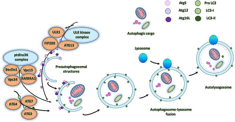
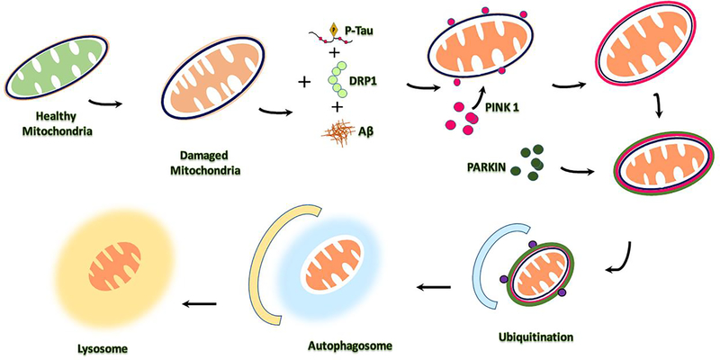
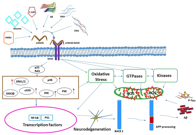
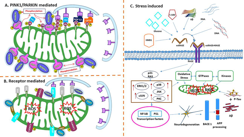
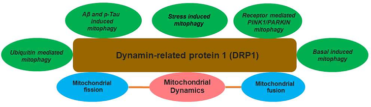
Alzheimer's disease (AD) is a progressive, mental illness without cure. Several years of intense research on postmortem AD brains, cell and mouse models of AD have revealed that multiple cellular changes are involved in the disease process, including mitochondrial abnormalities, synaptic damage, and glial/astrocytic activation, in addition to age-dependent accumulation of amyloid beta (Aβ) and hyperphosphorylated tau (p-tau). Synaptic damage and mitochondrial dysfunction are early cellular changes in the disease process. Healthy and functionally active mitochondria are essential for cellular functioning. Dysfunctional mitochondria play a central role in aging and AD. Mitophagy is a cellular process whereby damaged mitochondria are selectively removed from cell and mitochondrial quality and biogenesis. Mitophagy impairments cause the progressive accumulation of defective organelle and damaged mitochondria in cells. In AD, increased levels of Aβ and p-tau can induce reactive oxygen species (ROS) production, causing excessive fragmentation of mitochondria and promoting defective mitophagy. The current article discusses the latest developments of mitochondrial research and also highlights multiple types of mitophagy, including Aβ and p-tau-induced mitophagy, stress-induced mitophagy, receptor-mediated mitophagy, ubiquitin mediated mitophagy and basal mitophagy. This article also discusses the physiological states of mitochondria, including fission-fusion balance, Ca2+ transport, and mitochondrial transport in normal and diseased conditions. Our article summarizes current therapeutic interventions, like chemical or natural mitophagy enhancers, that influence mitophagy in AD. Our article discusses whether a partial reduction of Drp1 can be a mitophagy enhancer and a therapeutic target for mitophagy in AD and other neurological diseases.

Keywords: Alzheimer’s disease; Amyloid beta; Mitochondrial dysfunction; Mitophagy; Phosphorylated tau.


Conflict of interest statement

Conflicts of Interest: The authors declare no conflict of interest.

Figures

Figure 1.
Figure 1.. Molecular mechanism underlying autophagosome formation in normal cells.
The ULK complex consisting of ULK1/2–Atg13–FIP200–Atg101 is responsible for first initiation of autophagy, in response to certain signals. Formation of double-layered, membrane within the cytosol requires the PI3KC3 complex with contains Vps15–Vps34, Beclin-1, AMBRA1, and NBRF2. Next conjugation system is Atg, which add to the Atg5–Atg12–Atg16L complex, and LC3 conjugate system, which contains the Atg4, Atg7, Atg 3 to the elongating membrane along with the Pro LC3, LC3–1 and LC3–2. The membrane grows to enwrap a portion of the cytosol, forming an autophagosome. In the final step of the process, lysosomes fuse with the autophagosome, releasing lysosomal hydrolases into the interior, resulting in degradation of the vesicle contents.
Figure 2.
Figure 2.. A schematic illustrating of mitochondrial mitophagy events and mitochondria axonal transport.
(A) Microtubule-based transport of healthy mitochondria in neurons transfer from nucleus to cell soma (+) anterograde transportation through highlight interacting partners of kinesin-1-MIRO/TARK in three functional aspects. MIRO is the mediated by PINK-1/parkin and could be Ca2+ efflux is key for the transportation, which is mediated by the mitochondrial encounter structure (ERMES). (B) This attenuates mitochondrial mobility with Aβ and p-tau interactions foreword to the mitophagy of damaged mitochondria with abnormal mitochondrial dynamics. (C) Milton/TRAK and dynein mediates the retrograde axonal and dendritic transport. MIRO/TARK complex found to shown to be part of the ER- found at ERMES mitochondrial contact sites and showing Ca2+ exchange regulation in mitochondrial transport mechanism.
Figure 3.
Figure 3.
A brief autophagy and mitophagy events in Alzheimer’s disease with key toxic players like Aβ and p-tau and Drp1 inducers that progress the activation of PINK1/parkin mediated abnormal mitochondrial dynamics.
Figure 4.
Figure 4.. The extracellular and intracellular effects of AGEs induced ROS mechanism in AD.
The receptor for advanced glycation end products (RAGE) is a transmembrane, immunoglobulin-like receptor that exists and binds to ligands like HMB1, Glucose, S100, Aβ peptide, RNA, DNA. Ligand molecules binding at the extracellular domain of eRAGE initiates a complex with RAGE to form complex with esRAGE-RAGE and intracellular signaling cascade p21 RAS, stimulating NAD(P)H oxidase resulting in the production of reactive oxygen species (ROS). Up-regulation followed by ERK1/2, c-JUN, p38, JNK, PCK, GSK3β cellular events, and/or apoptosis with concomitant up regulation of and activation of NF-kB and PS1. The processing of APP has mainly focused on the correlation between RAGE activity and pathological conditions, such as BACE1 processing and Aβ accumulation and p-tau interaction in neuronal cells. The ROS formation influenced oxidative damage NADPH leads increased GTPases and kinases regulation to mitochondria mitophagy events leads neurodegeneration in RAGE mediated ROS signaling.
Figure 5.
Figure 5.
(A) Mechanism of mitophagy with PINK1–parkin based. PINK1 recruited on outer mitochondria membrane (OMM) and promoting parkin recruitment. PINK1 phosphorylates and recruiting parkin and ubiquitinated several outer membrane components with activated phospho mechanism. Ubiquitinated chains are attached to the OMM and polymerized subsequently phosphorylated by PINK1 serving as signal for the autophagic machinery. Adaptor proteins (p62, OPTN, NDP52) recognize phosphorylated poly-Ub chains on mitochondrial proteins and initiate autophagosome formation through binding with LC3B. (B). Receptor-mediated mitophagy. Specialized receptors, like NIX, BNIP3 and FUNDC1, expressed on the OMM in response to different stimuli. These receptors directly interact with LC3 to mediate mitochondrial elimination. NIX and BNIP3 phosphorylation enhances their association with LC3. FUNDC1 phosphorylation status, regulating mitochondrial dynamics during hypoxia. Mitophagy receptors promote fission of damaged organelles through the recruitment of DRP1, Opa1on the mitochondrial surface. Only parkin-dependent ubiquitination of NIX and BNIP3 mediated mitophagy in hypoxic condition. (C). Stress-mediated mitophagy. Ligand molecules binding at the extracellular domain of receptor for advanced glycation end products (eRAGE) initiates a complex with RAGE to form complex with esRAGE-RAGE and intracellular signaling cascade p21 RAS, stimulating NAD(P)H oxidase resulting the production of reactive oxygen species (ROS). Upregulated ERK1/2, c-JUN, p38, JNK, PCK, GSK3β cellular events, and/or apoptosis with concomitant up regulation of and activation of NF-kB and PS1. Processing of APP, RAGE activity and BACE1 processing leads Aβ accumulation and p-tau interaction resulting increased oxidative stress on mitochondria leads abnormal mitochondrial defects in AD.
Figure 6.
Figure 6.
Factors contributing mitophagy players in response to a variety of pathological stimuli connected to centric protein, dynamin related protein 1 of neuronal cellular changes in AD.

References

    1. Ahmad T, Aggarwal K, Pattnaik B, Mukherjee S, Sethi T, Tiwari BK, Kumar M, Micheal A, Mabalirajan U, Ghosh B, Sinha Roy S, Agrawal A 2013Computational classification of mitochondrial shapes reflects stress and redox state. Cell Death Dis 4:e461. - PMC - PubMed
    1. Ali S, McStay GP 2018. Regulation of Mitochondrial Dynamics by Proteolytic Processing and Protein Turnover. Antioxidants (Basel) 7, pii: E15. - PMC - PubMed
    1. Amakiri N, Kubosumi A, Tran J, Reddy PH 2019. Amyloid Beta and MicroRNAs in Alzheimer’s Disease. Front Neurosci. 13, 430. - PMC - PubMed
    1. Angelika BH, Hees JT 2019. Calcium Dysregulation and Mitochondrial Dysfunction Form A Vicious Cycle in Parkinson’s Disease. American Journal of Biomedical Science & Research 5, 246–249.
    1. Ashrafi G, Schwarz TL 2013. The pathways of mitophagy for quality control and clearance of mitochondria. Cell Death Differ, 20, 31–42. - PMC - PubMed

Publication types

Substances