Review
. 2020 Dec;594(23):4076-4084.
doi: 10.1002/1873-3468.13951. Epub 2020 Oct 20.

Mamma Mia, P-glycoprotein binds again

Affiliations
Review

Mamma Mia, P-glycoprotein binds again

Richard Callaghan et al. FEBS Lett. 2020 Dec.

Abstract

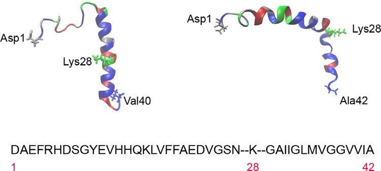
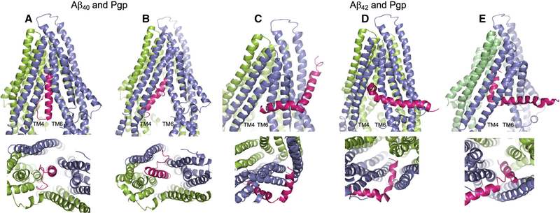
The levels of amyloid peptides in the brain are regulated by a clearance pathway from neurons to the blood-brain barrier. The first step is thought to involve diffusion from the plasma membrane to the interstitium. However, amyloid peptides are hydrophobic and avidly intercalate within membranes. The ABC transporter P-glycoprotein is implicated in the clearance of amyloid peptides across the blood-brain, but its role at neurons is undetermined. We here propose that P-glycoprotein mediates 'exit' of amyloid peptides from neurons. Indeed, amyloid peptides have physicochemical similarities to substrates of P-glycoprotein, but their larger size represents a conundrum. This review probes the plausibility of a mechanism for amyloid peptide transport by P-glycoprotein exploiting evolving biochemical and structural models.

Keywords: ABCB1; Alzheimer’s disease; MDR1; Pgp; amyloid peptides; blood-brain barrier; hydrophobic peptides; membrane transport.


Figures

Fig. 1.
Fig. 1.
Amino acid sequence and structure of the Aβ40 and Aβ42 peptides. The single letter code has been used for the sequence, which is divided to highlight lysine 28 for illustrative purposes. Residues have been colour-coded by type for Aβ40 (top left) and Aβ42 (top right). Hydrophobic amino acids are in blue, polar uncharged in red, acidic in silver and basic in green. Structures were obtained from PDB1iyt (Aβ42) and PDB1ba4 (Aβ40).
Fig. 2.
Fig. 2.
Docking of the Aβ40 and Aβ42 peptides to Pgp. The server CLUSPRO 2.0 was used to dock amyloid peptides Aβ40 (PDB: 1BA4) and Aβ42 (PDB: 1iYT) to Pgp (PDB: 4m1m). (A–E) Are side (upper) and bottom (lower) views of either peptide (hot pink) docked to Pgp (slate blue and chartreuse green). Pgp is represented with the domain swapped TM helices 4 and 5 (blue) shown at the left of each upper panel; and the opposite domain swapped TMs 10 and 11 (green) at the back right of each side view panel. (A) Aβ40 docked to Pgp with the nonpolar, helical end of the peptide uppermost in the Pgp cavity; and (B) Aβ40 docked to Pgp with the polar, random coil end of the peptide uppermost in the Pgp cavity. (C–E) Aβ42 docked to Pgp with the peptide at different stages of docking with Pgp. In each of the three docked modes, Aβ42 engages Pgp in the same orientation. (C) The short, nonpolar, C-terminal helix at the Pgp ‘gate’ between TM4 and TM6; (D) the Aβ42 helix within the Pgp cavity and at right angles to the Pgp TM helices; and (E) the helix wholly within the Pgp cavity and parallel to the Pgp TM helices. The figure was rendered with PYMOL (Molecular Graphics System, v2.4.0 Schrodinger). Standard settings were used, and no additional features such as attraction or repulsion were used. Scores for all docking modes were almost identical.

References

    1. Hardy J and Selkoe DJ (2002) The amyloid hypothesis of Alzheimer’s disease: progress and problems on the road to therapeutics. Science 297, 353–356. - PubMed
    1. Lam FC, Liu R, Lu P, Shapiro AB, Renoir JM, Sharom FJ and Reiner PB (2001) beta-Amyloid efflux mediated by p-glycoprotein. J Neurochem 76, 1121–1128. - PubMed
    1. Cirrito JR, Deane R, Fagan AM, Spinner ML, Parsadanian M, Finn MB, Jiang H, Prior JL, Sagare A, Bales KR et al., (2005) P-glycoprotein deficiency at the blood-brain barrier increases amyloid-beta deposition in an Alzheimer disease mouse model. J Clin Invest 115, 3285–3290. - PMC - PubMed
    1. Hartz AM, Miller DS and Bauer B (2010) Restoring blood-brain barrier P-glycoprotein reduces brain amyloid-beta in a mouse model of Alzheimer’s disease. Mol Pharmacol 77, 715–723. - PMC - PubMed
    1. Hartz AM, Zhong Y, Wolf A, LeVine H 3rd, Miller DS and Bauer B (2016) A beta40 reduces P-glycoprotein at the blood-brain barrier through the ubiquitin-proteasome pathway. J Neurosci 36, 1930–1941. - PMC - PubMed

Publication types

MeSH terms

Substances