. 2020 Oct 3;25(19):4534.
doi: 10.3390/molecules25194534.

New Sustainable Process for Hesperidin Isolation and Anti-Ageing Effects of Hesperidin Nanocrystals

Affiliations

New Sustainable Process for Hesperidin Isolation and Anti-Ageing Effects of Hesperidin Nanocrystals

Danijela Stanisic et al. Molecules. .

Abstract

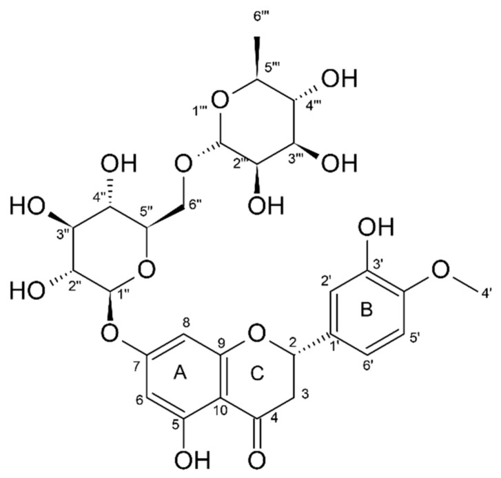
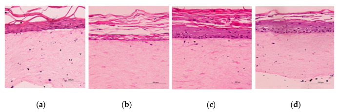
Hesperidin, a secondary orange (Citrus sinensis) metabolite, was extracted from orange bagasse. No organic solvents or additional energy consumption were used in the clean and sustainable process. Hesperidin purity was approximately 98% and had a yield of 1%. Hesperidin is a known supplement due to antioxidant, chelating, and anti-ageing properties. Herein, hesperidin application to eliminate dark eye circles, which are sensitive and thin skin regions, was studied. In addition, the proposed method for its aqueous extraction was especially important for human consumption. Further, the most effective methods for hesperidin nanonization were explored, after which the nanoemulsions were incorporated into a cream formulation that was formulated for a tropical climate. Silky cream formulations (oil in water) were tested in vitro on artificial 3D skin from cultured cells extracted from skin residues after plastic surgery. The proposed in vitro assay avoided tests of the different formulations in human volunteers and animals. It was shown that one of the nanonized hesperidin formulations was the most skin-friendly and might be used in cosmetics.

Keywords: anti-ageing; antioxidant; hesperidin; nanocrystals; new-clean process extraction.


Conflict of interest statement

The authors declare no conflict of interest.

Figures

Figure 1
Figure 1
Chemical structure of hesperidin (2S)-5-hydroxy-2-(3-hydroxy-4-methoxyphenyl)-7-[(2S,3R,4S,5S,6R)-3,4,5-trihydroxy-6-[[(2R,3R,4R,5R,6S)-3,4,5-trihydroxy-6-methyloxan-2-yl]oxymethyl]oxan-2-yl]oxy-2,3-dihydrochromen-4-one). HSD carbon atoms are numbered, and A–C rings are shown.
Figure 2
Figure 2
Fourier transform infrared (FT-IR) spectra of hesperidin (HSD, dotted line), copper(II) acetate (Cu(II), dashed line), and HSD–Cu(II) complex (full line) on a Cary 630 FT-IR spectrometer equipped with a monolithic design diamond Attenuated Total Reflectance (ATR) accessory (Agilent Technologies Inc.). Hesperidin is a chelating agent of metal multivalent ions (Me2+). The creation of the complex involves -OH groups in ortho- positions that coordinate with divalent cations [1].
Figure 3
Figure 3
Illustration of fluorescence spectroscopy data: (a) Collagenase (Clostridium hystolicum) at 37 °C upon addition of HSD (in DMSO) at concentrations from 0.08 to 0.90 mmol L−1. (b) Diagram of log((F0−F)/F) in function of log[HSD].
Figure 4
Figure 4
Scanning electron micrographs (left; scale bar = 500 nm) of formulation I and transmission electron micrographs (right; scale bar = 200 nm) of formulation I. Scanning electron micrographs of formulation III (left; scale bar = 1 µm) and formulation IV (right; scale bar = 500 nm), respectively. Scanning electron micrographs of formulation V (left; scale bar = 1 µm) and atomic force micrographs of formulation VI (right; scale bar = 4 µm). Scanning electron micrographs of formulations VII and VIII (left; scale bar = 2 µm).
Figure 5
Figure 5
Photomicrographs of Histological Analysis—Eosin stain (sample in paraffin) of the full thickness skin (FTS) equivalent skin model after the cream application assay (a) negative control: sodium chloride 0.9%; (b) positive control: HSD-DMSO, (c) A1 cream, and (d) A2 cream. The skin models were generated using primary human keratinocytes and fibroblasts—the FTS model; (Scale bar = 500 µm).

References

    1. Tasic L., Mandic B., Barros C.H.N., Cypriano D.Z., Stanisic D., Schultz L.G., da Silva L.L., Mariño M.A.M., Queiroz V.L. In: Citrus Fruits: Productions, Consumption and Health Benefits. Simons D., editor. Nova Science Publishers; New York, NY, USA: 2016. pp. 27–70. Chapter 2.
    1. Stanisic D., Costa A.F., Cruz G., Durán N., Tasic L. In: Studies in Natural Products Chemistry. Rahman A.-U., editor. Volume 58. Elsevier Science; Amsterdam, The Netherlands: 2018. pp. 161–210. Chapter 6.
    1. Ratz-Lyko A., Arct J., Majewski S., Pytkowska K. Influence of polyphenols on the physiological processes in the skin. Phytother. Res. 2015;29:509–517. doi: 10.1002/ptr.5289. - DOI - PubMed
    1. Chanet A., Milenkovic D., Manach C., Mazur A., Morand C. Citrus Flavanones: What Is Their Role in Cardiovascular Protection? J. Agric. Food Chem. 2012;60:8809–8822. doi: 10.1021/jf300669s. - DOI - PubMed
    1. Garg A., Garg S., Zaneveld L.J.D., Singla A.K. Chemistry and pharmacology of the citrus bioflavonoid Hsd. Phytother. Res. 2001;15:655–669. doi: 10.1002/ptr.1074. - DOI - PubMed