Review
. 2020 Oct 3;25(19):4537.
doi: 10.3390/molecules25194537.

Glucosinolates: Natural Occurrence, Biosynthesis, Accessibility, Isolation, Structures, and Biological Activities

Affiliations
Review

Glucosinolates: Natural Occurrence, Biosynthesis, Accessibility, Isolation, Structures, and Biological Activities

V P Thinh Nguyen et al. Molecules. .

Abstract

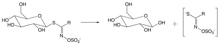
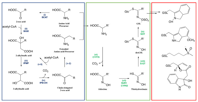
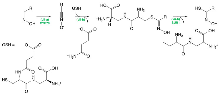
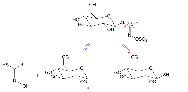
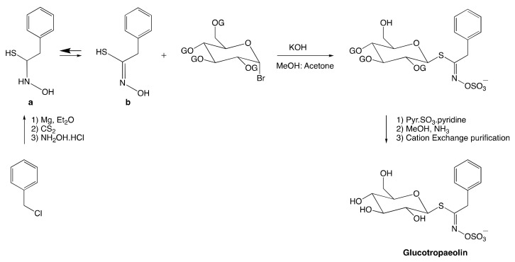
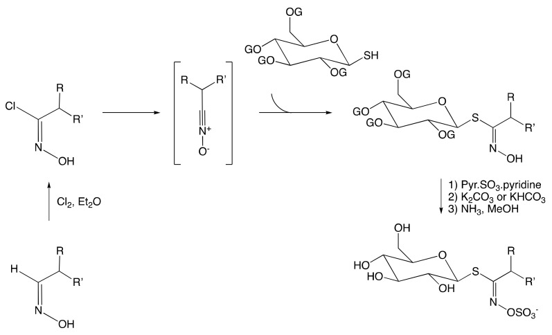
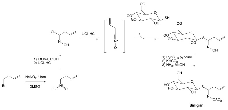
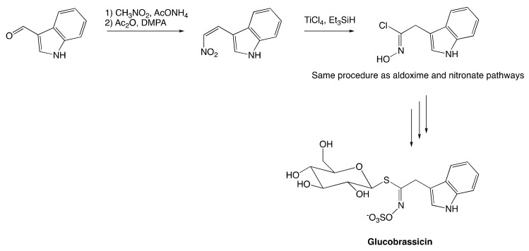
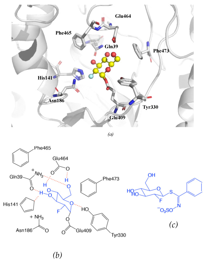
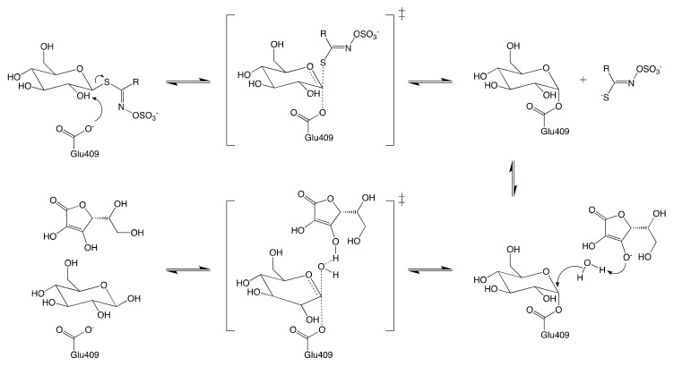
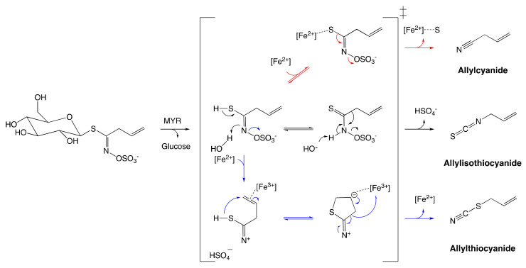
Glucosinolates (GSLs) are secondary plant metabolites abundantly found in plant order Brassicales. GSLs are constituted by an S-β-d-glucopyrano unit anomerically connected to O-sulfated (Z)-thiohydroximate moiety. The side-chain of the O-sulfate thiohydroximate moiety, which is derived from a different amino acid, contributes to the diversity of natural GSL, with more than 130 structures identified and validated to this day. Both the structural diversity of GSL and their biological implication in plants have been biochemically studied. Although chemical syntheses of GSL have been devised to give access to these secondary metabolites, direct extraction from biomass remains the conventional method to isolate natural GSL. While intact GSLs are biologically inactive, various products, including isothiocyanates, nitriles, epithionitriles, and cyanides obtained through their hydrolysis of GSLs, exhibit many different biological activities, among which several therapeutic benefits have been suggested. This article reviews natural occurrence, accessibility via chemical, synthetic biochemical pathways of GSL, and the current methodology of extraction, purification, and characterization. Structural information, including the most recent classification of GSL, and their stability and storage conditions will also be discussed. The biological perspective will also be explored to demonstrate the importance of these prominent metabolites.

Keywords: Brassicaceae family; Brassicales; Moringacea family; glucosinolates; myrosinases.


Conflict of interest statement

The authors declare no conflict of interest.

Figures

Figure 1
Figure 1
Hydrolysis of glucosinolate (GSL) by myrosinase (MYR) upon tissue disruption. (R = alkyl, aryl, indole).
Figure 2
Figure 2
Three separate phases of glucosinolate biosynthesis: R indicates the variable amino acid precursors, and R’ indicates either original or extended amino acid. The blue box indicates the chain elongation phase, the green box indicates the reconfiguration phase yielding the core structure of glucosinolate, and the red box indicates the glucosinolate side-chain modification phase of the glucosinolate core structure with some examples from Table 2. The figure was adapted from the biosynthesis of GSL proposed by Graser et al. [46].
Figure 3
Figure 3
Conversion of Aldoximes to Thiohydroximic Acids. GSH: Glutathione.
Figure 4
Figure 4
Retrosynthesis approach to GSL synthesis: anomeric disconnection (blue), hydroximate disconnection (red). OG: suitable protecting group.
Figure 5
Figure 5
Synthesis of glucotropaeolin. OG represents a suitable protecting group.
Figure 6
Figure 6
Synthesis of GSL following the aldoxime pathway (R, R’ = H, alkyl, or aryl).
Figure 7
Figure 7
Synthesis of sinigrin employing nitronate pathway.
Figure 8
Figure 8
Synthesis of glucobrassicin.
Figure 9
Figure 9
Overview of the active site of Sinapis alba Myrosinase showing interactions between residues and the 2-deoxy-2-fluoroglucosinolate (2FG) as substrate (Protein Data Bank accession number 1E70, resolution: 1.65 Å) [172]. Red dashed lines show hydrogen bonding interactions between the substrate and MYR residues within the active site. (a) Representation of the active site of Sinapis alba Myrosinase generated using PyMol. (b) Chemical structure representation of the MYR-2FG. (c) Structure of 2-deoxy-2-fluoroglucosinolate.
Figure 10
Figure 10
Schematic reaction mechanism of MYR in the presence of ascorbic acid.
Figure 11
Figure 11
Reconfiguration of unstable allylglucosinolate aglucone upon myrosinase-catalyzed hydrolysis. The black arrow pathway shows the formation of allylisothiocyanates employing spontaneous Lossen arrangement. The Blue arrow pathway shows the formation of allylthiocyanate assisted by protein specifier. The red arrow pathway indicates the formation of allylcyanide assisted by protein specifier. The figure was adapted from Eisenschmidt-Bönn et al. [179].

References

    1. Wittstock U., Halkier B.A. Cytochrome P450 CYP79A2 from Arabidopsis thaliana L. catalyzes the conversion of L-phenylalanine to phenylacetaldoxime in the biosynthesis of benzylglucosinolate. J. Biol. Chem. 2000;275:14659–14666. doi: 10.1074/jbc.275.19.14659. - DOI - PubMed
    1. Borgen B.H., Thangstad O.P., Ahuja I., Rossiter J.T., Bones A.M. Removing the mustard oil bomb from seeds: Transgenic ablation of myrosin cells in oilseed rape (Brassica napus) produces MINELESS seeds. J. Exp. Bot. 2010;61:1683–1697. doi: 10.1093/jxb/erq039. - DOI - PMC - PubMed
    1. Martínez-Ballesta M., Moreno-Fernández D.A., Castejón D., Ochando C., Morandini P.A., Carvajal M. The impact of the absence of aliphatic glucosinolates on water transport under salt stress in Arabidopsis thaliana. Front. Plant Sci. 2015;6:1–12. doi: 10.3389/fpls.2015.00524. - DOI - PMC - PubMed
    1. Blaževic I., Montaut S., Burcul F., Olsen C.E., Burow M., Rollin P., Agerbirk N. Glucosinolate structural diversity, identification, chemical synthesis and metabolism in plants. Phytochemistry. 2020;169:112100. doi: 10.1016/j.phytochem.2019.112100. - DOI - PubMed
    1. Borek V., Morra M.J., Brown P.D., McCaffrey J.P. Allelochemicals Produced during Sinigrin Decomposition in Soil. J. Agric. Food Chem. 1994;42:1030–1034. doi: 10.1021/jf00040a037. - DOI

LinkOut - more resources