Review
. 2020 Oct 3;21(19):7307.
doi: 10.3390/ijms21197307.

The Immune Microenvironment in Pancreatic Cancer

Affiliations
Review

The Immune Microenvironment in Pancreatic Cancer

Magdalena Huber et al. Int J Mol Sci. .

Abstract

The biology of solid tumors is strongly determined by the interactions of cancer cells with their surrounding microenvironment. In this regard, pancreatic cancer (pancreatic ductal adenocarcinoma, PDAC) represents a paradigmatic example for the multitude of possible tumor-stroma interactions. PDAC has proven particularly refractory to novel immunotherapies, which is a fact that is mediated by a unique assemblage of various immune cells creating a strongly immunosuppressive environment in which this cancer type thrives. In this review, we outline currently available knowledge on the cross-talk between tumor cells and the cellular immune microenvironment, highlighting the physiological and pathological cellular interactions, as well as the resulting therapeutic approaches derived thereof. Hopefully a better understanding of the complex tumor-stroma interactions will one day lead to a significant advancement in patient care.

Keywords: T-cells; cancer-associated fibroblasts; immunotherapy; macrophages; natural killer cells; neutrophils; pancreatic cancer; tumor stroma.


Conflict of interest statement

The authors declare no conflict of interest.

Figures

Figure 1
Figure 1
Tumor-associated macrophages (TAMs) and myeloid-derived suppressor cells (MDSCs) in pancreatic ductal adenocarcinoma (PDAC). (A) Immunohistochemical staining of pan-monocytic marker CD68 (macrosialin; dark brown signals) in examples of well-differentiated, stroma-rich (upper panel) and poorly differentiated, stroma-poor (lower panel) pancreatic ductal adenocarcinoma. Scale bar: 100 µm. (B) Scheme of TAM functions in PDAC.
Figure 2
Figure 2
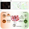
Natural killer cells and their roles in PDAC. (A) Immunohistochemical staining of CD56 reveals disseminated natural killer (NK)/NKT (Natural killer T-cells) cells (brown). The depicted case shows a rather high density of NK cells considering the distinct heterogeneity of NK cell infiltration, which is generally low in PDAC tissue. (B) PDAC NK cells isolated from the periphery are characterized by a reduced expression of cytotoxicity receptors and exhibit impaired anti-tumor activity, which is induced by mediators of the tumor microenvironment. Restoration of NK cell functions is possible, e.g., via ex vivo activation with IL-2 and gamma-irradiated feeder cell lines. The phenotype of tumor-infiltrating NK cells or other innate lymphocytes such as ILC1 cells and their activity is poorly understood. Gzm: granzyme; Prf: perforin.
Figure 3
Figure 3
Tumor-associated neutrophils in pancreatic cancer. (A) Neutrophils in PDAC tumor stroma, stained with an antibody against Myeloperoxidase (MPO, pink). Ductal structures with tumor cells are stained in brown. Scale bar: 200 µm. (B) Signaling events leading to neutrophil recruitment (left side) and neutrophil polarization (right side). Activating signals are indicated by green, and suppressing signals by red arrows. Note that neutrophils can activate macrophages and dendritic cells, while they suppress lymphocytes. When polarized, the tumor-associated neutrophil (TAN) N1 type can activate CD8-positive T cells, whereas TAN N2 secretes a cocktail of tumor-promoting factors, such as hepatocyte growth factor (HGF), matrix metalloproteinase (MMPs), and neutrophils specific protease neutrophil elastase (NE). Furthermore, TAN N2 cells can cause an increased production of reactive oxygen and nitrogen species (ROS and RNS, respectively) and can cause hypoxic signaling.
Figure 4
Figure 4
Role of T cell subpopulations in PDAC. (A) Compound image of a pancreatic cancer tissue microarray (TMA) section (blue: CD3; yellow: CD8; red: programmed cell death 1 (PD-1); orange: PanCK). (B) Phenotype map, same section as in A (yellow: CD8+ PD-1- T cells; orange: CD8+ PD-1+ T cells; purple: epithelium/tumor; light blue: CD8- T cells; green: everything else). Magnified area is densely infiltrated by PD-1neg CD8+ T cells (CTLs), as well as PD-1pos CD8+ CTLs. Remnants of epithelial/tumor cells are found in close vicinity to exhausted PD-1pos T cells. (C) Th2 cells expressing the transcription factor GATA3 can be induced by (i) interleukin (IL)-1α and IL-1β primed cancer-associated fibroblasts (CAFs) producing thymic stromal lymphopoietin (TSLP), which diverts dendritic cells (DCs) towards the Th2 inducing phenotype; (ii) B cells and tumor-associated macrophages in a Bruton tyrosine kinase (BTK)- and Pi3Kg-dependent manner; and (iii) by the microbiome via TLR-signaling. Th2 cells recruit tumor-promoting macrophages (M2) and can directly induce tumor cell proliferation via IL-4. Oncogenic Kras mediates Th17 cell recruitment into the tumor microenvironment. Via secreted IL-17 and IL-21, Th17 cells enhance early PDAC progression. An alternative p38 mitogen-activated protein kinase (MAPK) activation pathway in CD4+ T cells directs Th17 cell differentiation. Beneficial CTL and Th1 responses in PDAC are suppressed by Tregs via the immunomodulation of CD11c+ DCs. Tregs can be beneficial in PDAC as they suppress tumor progression via the induction of myofibroblastic CAF (myCAFs) by secreted transforming growth factor beta (TGFβ). CTL and Th1 cells restrict PDAC growth via secreted IFN-γ and direct cytotoxicity. Th1 promotes CTL responses and M1 differentiation, thereby restricting tumor growth. CTL accumulation in the tumor microenvironment can be induced by cross-presenting CD103+CD11c+ DCs, while tumor cells, via CXCL1-dependent attracting myeloid cells, restrict CD8+ T cell accumulation in tumors. High-quality neoantigens, including MUC16, contribute to long-term survival in PDAC.
Figure 5
Figure 5
The functions of CAFs in the immune landscape of PDAC. (A) Immunohistochemical example of tumor cell localization (CA19-9 staining in brown, upper panel) and αSMA-positive myCAFs (also brown, lower panel) in human PDAC. Note that the myCAFs are in close vicinity to the tumor cells. (B) Tumor cells polarize quiescent pancreatic stellate cells towards different subtypes of cancer-associated fibroblasts (CAFs). Contractile myCAFs and inflammatory-CAFs (iCAFs) represent the two major populations. IL1-induced iCAFs secrete a large number of soluble mediators that affect a plethora of immune cells, essentially generating an immunosuppressive environment favoring tumor cell growth. In addition, immune cells might also be suppressed by CAF-mediated metabolic competition and direct cell-cell contact (not shown).
Figure 6
Figure 6
Cross-talk between cancer cells and immune cells mediating immunotherapeutic approaches in pancreatic cancer patients. APC, antigen-presenting cell; CAR, chimeric antigen receptor; CTL, cytotoxic T cell; LAG-3, lymphocyte-activation gene-3; PD-1, programmed death receptor-1; PD-L1, programmed death-ligand 1; Tex, exhausted T cell.

References

    1. Siegel R.L., Miller K.D., Jemal A. Cancer statistics, 2020. CA Cancer J. Clin. 2020;70:7–30. doi: 10.3322/caac.21590. - DOI - PubMed
    1. Rahib L., Smith B.D., Aizenberg R., Rosenzweig A.B., Fleshman J.M., Matrisian L. Projecting Cancer Incidence and Deaths to 2030: The Unexpected Burden of Thyroid, Liver, and Pancreas Cancers in the United States. Cancer Res. 2014;74:2913–2921. doi: 10.1158/0008-5472.CAN-14-0155. - DOI - PubMed
    1. Singhi A.D., Koay E.J., Chari S.T., Maitra A. Early Detection of Pancreatic Cancer: Opportunities and Challenges. Gastroenterology. 2019;156:2024–2040. doi: 10.1053/j.gastro.2019.01.259. - DOI - PMC - PubMed
    1. Strobel O., Neoptolemos J., Jäger D., Büchler M.W. Optimizing the outcomes of pancreatic cancer surgery. Nat. Rev. Clin. Oncol. 2018;16:11–26. doi: 10.1038/s41571-018-0112-1. - DOI - PubMed
    1. Versteijne E., Suker M., Groothuis K., Akkermans-Vogelaar J.M., Besselink M.G., Bonsing B.A., Buijsen J., Busch O.R., Creemers G.-J.M., Van Dam R.M., et al. Preoperative Chemoradiotherapy Versus Immediate Surgery for Resectable and Borderline Resectable Pancreatic Cancer: Results of the Dutch Randomized Phase III PREOPANC Trial. J. Clin. Oncol. 2020;38:1763–1773. doi: 10.1200/JCO.19.02274. - DOI - PMC - PubMed

MeSH terms

LinkOut - more resources