Review
. 2020 Oct 2;21(19):7290.
doi: 10.3390/ijms21197290.

Retinal Degeneration and Alzheimer's Disease: An Evolving Link

Affiliations
Review

Retinal Degeneration and Alzheimer's Disease: An Evolving Link

Ajay Ashok et al. Int J Mol Sci. .

Abstract

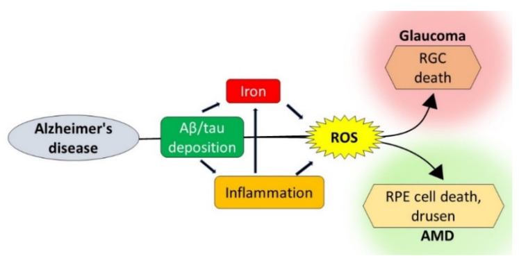
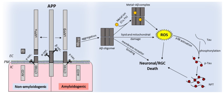
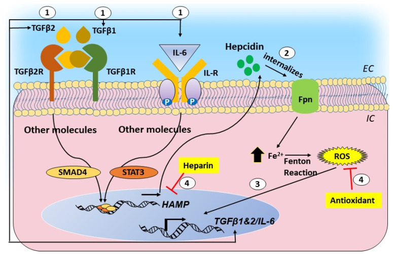
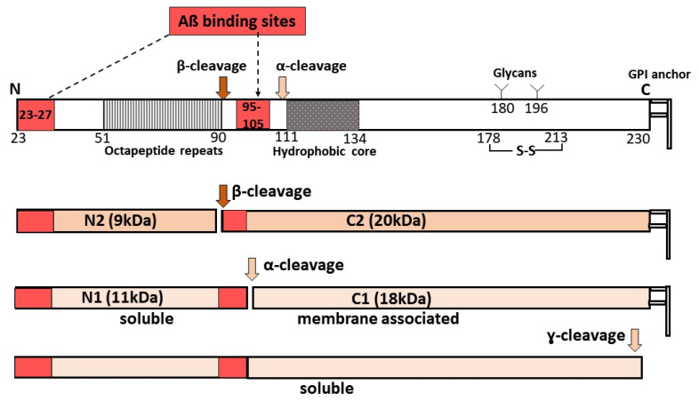
Age-related macular degeneration (AMD) and glaucoma are degenerative conditions of the retina and a significant cause of irreversible blindness in developed countries. Alzheimer's disease (AD), the most common dementia of the elderly, is often associated with AMD and glaucoma. The cardinal features of AD include extracellular accumulation of amyloid β (Aβ) and intracellular deposits of hyper-phosphorylated tau (p-tau). Neuroinflammation and brain iron dyshomeostasis accompany Aβ and p-tau deposits and, together, lead to progressive neuronal death and dementia. The accumulation of Aβ and iron in drusen, the hallmark of AMD, and Aβ and p-tau in retinal ganglion cells (RGC), the main retinal cell type implicated in glaucoma, and accompanying inflammation suggest overlapping pathology. Visual abnormalities are prominent in AD and are believed to develop before cognitive decline. Some are caused by degeneration of the visual cortex, while others are due to RGC loss or AMD-associated retinal degeneration. Here, we review recent information on Aβ, p-tau, chronic inflammation, and iron dyshomeostasis as common pathogenic mechanisms linking the three degenerative conditions, and iron chelation as a common therapeutic option for these disorders. Additionally discussed is the role of prion protein, infamous for prion disorders, in Aβ-mediated toxicity and, paradoxically, in neuroprotection.

Keywords: Alzheimer’s disease; age related macular degeneration; drusen; glaucoma; inflammation; iron; oxidative stress; prion protein; reactive oxygen species; retinal degeneration.


Conflict of interest statement

The authors declare no conflict of interest.

Figures

Figure 1
Figure 1
Pathogenic pathways shared by Alzheimer’s disease (AD), AMD, and glaucoma: AD-associated amyloid β (Aβ) and tau deposits lead to inflammation and iron accumulation, increasing ROS. Aβ, hyper-phosphorylated tau (p-tau), inflammation, and ROS together lead to RGC death typical of glaucoma and retinal pigment epithelial (RPE) dysfunction associated with AMD. ROS: reactive oxygen species, RGC: retinal ganglion cells, and AMD: age-related macular degeneration.
Figure 2
Figure 2
Schematic representation of nonamyloidogenic and amyloidogenic processing of the amyloid precursor protein (APP): nonamyloidogenic processing of APP is mediated by α-secretase followed by γ-secretase, releasing soluble APPα and intracellular fragments AICD and P3. Amyloidogenic processing involves cleavage by β-secretase followed by γ-secretase, which releases toxic Aβ1−42 [79,80]. Aβ can oligomerize and form cytotoxic metal-Aβ complexes that generate ROS, disrupt the lipid bilayer, compromise mitochondrial function, or initiate aberrant signaling cascades. Activation of p38 by ROS phosphorylates tau results in NFTs. IC: intracellular, EC: extracellular, NFTs: neurofibrillary tangles, and PM: plasma membrane.
Figure 3
Figure 3
Graphical representation of the cytokine-hepcidin-iron feed-forward loop and its disruption by hepcidin antagonists and antioxidants: (1) transforming growth factor beta 1 and 2 (TGF-β1 and 2) and interleukin (IL)-6 upregulate hepcidin through the SMAD and signal transducer and activator of transcription (STAT)-mediated pathways. (2) Hepcidin causes the downregulation of ferroportin and intracellular accumulation of iron. (3) Iron-catalyzed ROS promotes the transcriptional activation of TGF-β1 and 2 and IL-6, creating a self-sustained feed-forward loop. (4) Hepcidin antagonists and antioxidants disrupt this loop.
Figure 4
Figure 4
Processing of prion protein (PrPC) and Aβ binding: PrPC undergoes α-, β-, or γ-cleavage, releasing extracellular N-terminal fragments N1 (α-cleavage), N2 (β-cleavage), or soluble PrPC. Full-length PrPC has two Aβ binding sites. N2 has one, and N1 and soluble PrPC have two Aβ binding sites that could sequester soluble Aβ. The truncated C2 and C1 are attached to the plasma membrane, and the presence of one Aβ-binding site on C2 is likely to transmit the toxic signal of Aβ.

References

    1. Kent S.A., Spires-Jones T.L., Durrant C.S. The physiological roles of tau and Abeta: Implications for Alzheimer’s disease pathology and therapeutics. Acta Neuropathol. 2020;140:1–31. doi: 10.1007/s00401-020-02196-w. - DOI - PMC - PubMed
    1. Rajmohan R., Reddy P.H. Amyloid-β and Phosphorylated Tau Accumulations Cause Abnormalities at Synapses of Alzheimer’s disease Neurons. J. Alzheimers Dis. 2017;57:975–999. doi: 10.3233/JAD-160612. - DOI - PMC - PubMed
    1. Kinney J.W., Bemiller S.M., Murtishaw A.S., Leisgang A.M., Salazar A.M., Lamb B.T. Inflammation as a central mechanism in Alzheimer’s disease. Alzheimers Dement. 2018;4:575–590. doi: 10.1016/j.trci.2018.06.014. - DOI - PMC - PubMed
    1. Cheignon C., Tomas M., Bonnefont-Rousselot D., Faller P., Hureau C., Collin F. Oxidative stress and the amyloid β peptide in Alzheimer’s disease. Redox. Biol. 2018;14:450–464. doi: 10.1016/j.redox.2017.10.014. - DOI - PMC - PubMed
    1. Collin F., Cheignon C., Hureau C. Oxidative stress as a biomarker for Alzheimer’s disease. Future Med. 2018;12:201–203. doi: 10.2217/bmm-2017-0456. - DOI - PubMed

MeSH terms