

Syndromic Inherited Retinal Diseases: Genetic, Clinical and Diagnostic Aspects
- PMID: 33023209
- PMCID: PMC7600643
- DOI: 10.3390/diagnostics10100779
Syndromic Inherited Retinal Diseases: Genetic, Clinical and Diagnostic Aspects
Abstract
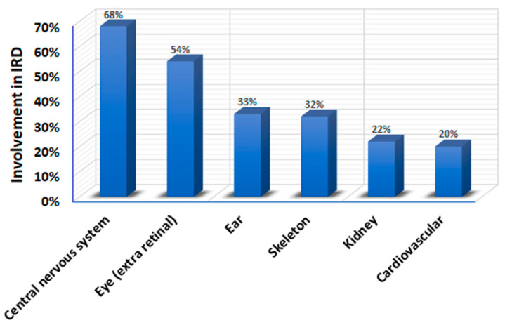
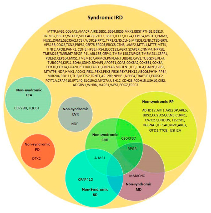
Inherited retinal diseases (IRDs), which are among the most common genetic diseases in humans, define a clinically and genetically heterogeneous group of disorders. Over 80 forms of syndromic IRDs have been described. Approximately 200 genes are associated with these syndromes. The majority of syndromic IRDs are recessively inherited and rare. Many, although not all, syndromic IRDs can be classified into one of two major disease groups: inborn errors of metabolism and ciliopathies. Besides the retina, the systems and organs most commonly involved in syndromic IRDs are the central nervous system, ophthalmic extra-retinal tissues, ear, skeleton, kidney and the cardiovascular system. Due to the high degree of phenotypic variability and phenotypic overlap found in syndromic IRDs, correct diagnosis based on phenotypic features alone may be challenging and sometimes misleading. Therefore, genetic testing has become the benchmark for the diagnosis and management of patients with these conditions, as it complements the clinical findings and facilitates an accurate clinical diagnosis and treatment.
Keywords: inherited retinal diseases; retina; syndrome.
Conflict of interest statement
The authors declare no conflict of interest. The funders had no role in the design of the study; in the collection, analyses, or interpretation of data; in the writing of the manuscript, or in the decision to publish the results.
Figures
References
-
- Thiadens A.A., Phan T.M., Zekveld-Vroon R.C., Leroy B.P., van den Born L.I., Hoyng C.B., Klaver C.C., Roosing S., Pott J.W., van Schooneveld M.J., et al. Clinical course, genetic etiology, and visual outcome in cone and cone-rod dystrophy. Ophthalmology. 2012;119:819–826. doi: 10.1016/j.ophtha.2011.10.011. - DOI - PubMed
-
- Tsang S.H., Sharma T. Rod Monochromatism (Achromatopsia) Adv. Exp. Med. Biol. 2018;1085:119–123. - PubMed
Publication types
LinkOut - more resources
Full Text Sources
