. 2021 Feb;27(1):121-128.
doi: 10.1177/1591019920963816. Epub 2020 Oct 6.

A new subtype of intracranial dural AVF according to the patterns of venous drainage

Affiliations

A new subtype of intracranial dural AVF according to the patterns of venous drainage

Alfredo Casasco et al. Interv Neuroradiol. 2021 Feb.

Abstract

Background and purpose: A well-known classification of dural arteriovenous fistulas (DAVFs) according to the patterns of venous drainage was described in 1977 by Djindjian, Merland et al. and later revised by Cognard, Merland et al. in 1995. They described 5 types of DAVFs assuming that the type of venous drainage is directly correlated with neurologic symptoms and in particular with hemorrhagic risk. We present a series of cases that combines type IV (DAVF with cortical venous drainage associated with venous ectasia) and type V (DAVF with spinal venous drainage), which we named type IV + V.

Materials and methods: A retrospective study between 2012 and 2020 in 2 Hospitals was performed on patients that met inclusion criteria for a diagnosis of this type of DAVF. Demographics, location, clinical presentation and outcomes of endovascular embolization were studied.

Results: Five (2,3%) patients out of 220 had a type IV + V DAVF. All cases had an aggressive presentation, either subarachnoid hemorrhage, myelopathy or both. All patients were treated with endovascular transarterial embolization achieving complete angiographic occlusion in one session and total remission of symptoms at 3 months.

Conclusions: This rare type of DAVF, combines two aggressive venous drainage patterns. For that reason, patients with type IV+V DAVF probably have a more aggressive natural history and worst outcome due to risk of intracranial and/or spinal hemorrhage and myelopathy, thus requiring urgent diagnostic and treatment. Larger studies are needed to better understand this type of DAVF.

Keywords: Intracranial hemorrhage; dural arteriovenous fistula; myelopathy.


Conflict of interest statement

Declaration of conflicting interests: The author(s) declared no potential conflicts of interest with respect to the research, authorship, and/or publication of this article.

Figures

Figure 1.
Figure 1.
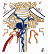
Anatomic sketch of a type IV + V dural fistula illustrating the association of venous ectasia (black arrow) with perimedular drainage (black triangle).
Figure 2.
Figure 2.
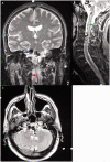
Patient 3. MRI (a) T2 coronal, (b) T2 sagittal and (c) T2 axial show intracranial venous ectasias (blue arrow) and descending anterior perimedular drainage (red arrows). Note the centromedular hypersignal (surrogate for retrograde venous hypertension) (green arrow).
Figure 3.
Figure 3.
Patient 3. (a) Right ICA angiography. Tentorial dural fistula with associated venous ectasia (blue arrow) irrigated by an intracavernous tentorial branch (red arrow). (b) Selective study of the right ECA showing the fistula supply through the petrosal branch of the MMAmiddle (red arrow), the retromastoid branch of the OA (green arrow) and the PAA (white arrow). The confluence of the feeders to the fistulous point and the primary venous drainage are well seen (black arrow). (c) Anterior perimedular drainage descends extremely dilated (red arrow). (d) Supraselective catheterization of the petrosal branch of the MMA showing the primary venous drainage of the fistula. (e) The treatment strategy was to perform the NBCA embolization from the ECA under temporary balloon occlusion of the ICA at the origin of the cavernous branches (microcatheter in the petrosal branch pointed with black arrows). F: NBCA cast showing the filling of the primary draining vein (blue arrow), part of the ectasia (black arrow) and retrograde filling of the tentorial branches of the ICA(white arrow). ICA: the internal carotid artery, ECA: external carotid artery, MMA: middle meningeal artery, OA: occipital artery, PAA: posterior auricular artery, NBCA: N-Butyl-2-cyanoacrylate
Figure 4.
Figure 4.
Patient 3. Follow up studies. (a) Right ICA angiogram showing complete occlusion of the type IV + V dural fistula at 3 months. (b) MRI 3 months post treatment. T2 sagittal. Absence of venous ectasias and descending perimedular drainage. Normalization of the hypersignal of the centromedular parenchyma.
Figure 5.
Figure 5.
Patient 2 Angiography: (a) Type IV + V DAVFof the right condylar foramen region. Selective right ECA (AP view)showing the supply from the APA (black arrow), which originates from a common occipitopharyngeal trunk (white arrow). Intracranial venous ectasia is identified (green arrow). (b,c) Late venous drainage (AP and Lateral view) showing the association of intracranial venous ectasias plus anterior (black arrow) and posterior (blue arrow) descending perimedular venous drainage. (d) Supraselective catheterization of the right ascending pharyngeal artery identifying the primary venous drainage of the fistula. (e) Post embolization Cast showing the occlusion of the fistulous point (white arrow) and the origin of the primary venous drainage. (f) ECA angiogram post-embolization showing the complete occlusion of the fistula. DAVF: Dural arteriovenous fistula, APA: ascending pharyngeal artery, AP: anteroposterior, ECA: external carotid artery.

References

    1. Newton TH, Weidner W, Greitz T. Involvement of the dural arteries in intra-cranial arteriovenous malformations. Radiology 1968; 90: 27–35. - PubMed
    1. Gandhi D, Chen J, Pearl M, et al. Intracranial dural arteriovenous fistulas: classification, imaging findings, and treatment. Am J Neuroradiol 2012; 33: 1007–1013. - PMC - PubMed
    1. Djindjian R, Merland JJ, Theron J. Superselective arteriography of the external carotid artery. New York, NY: Springer-Verlag, 1977, pp.606–628.
    1. Cognard C, Gobin YP, Pierot L, et al. Cerebral dural arteriovenous fistulas: clinical and angiographic correlation with a revised classification of venous drainage. Radiology 1995; 194: 671–680. - PubMed
    1. Gomez J, Amin AG, Gregg L, et al. Classification schemes of cranial dural arteriovenous fistulas. Neurosurg Clin N Am 2012; 23: 55–62. - PubMed

MeSH terms

LinkOut - more resources