Observational Study
. 2021 Feb;76(2):108-116.
doi: 10.1016/j.crad.2020.09.002. Epub 2020 Sep 15.

COVID-19-related intracranial imaging findings: a large single-centre experience

Affiliations
Observational Study

COVID-19-related intracranial imaging findings: a large single-centre experience

V Sawlani et al. Clin Radiol. 2021 Feb.

Abstract

Aim: To describe the neuroradiological changes in patients with coronavirus disease 2019 (COVID-19).

Materials and methods: A retrospective review was undertaken of 3,403 patients who were confirmed positive for severe acute respiratory syndrome coronavirus 2 (SARS-CoV-2) infection, and admitted to Queen Elizabeth Hospital Birmingham, University Hospitals Birmingham NHS Foundation Trust, Birmingham, UK between 1 March 2020 and 31 May 2020, and who underwent neuroimaging. Abnormal brain imaging was evaluated in detail and various imaging patterns on magnetic resonance imaging MRI were identified.

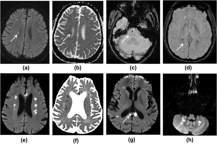
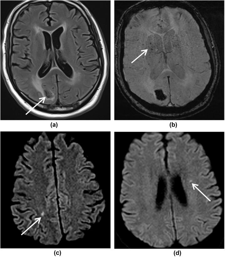
Results: Of the 3,403 patients with COVID-19, 167 (4.9%) had neurological signs or symptoms warranting neuroimaging. The most common indications were delirium (44/167, 26%), focal neurology (37/167, 22%), and altered consciousness (34/167, 20%). Neuroimaging showed abnormalities in 23% of patients, with MRI being abnormal in 20 patients and computed tomography (CT) in 18 patients. The most consistent neuroradiological finding was microhaemorrhage with a predilection for the splenium of the corpus callosum (12/20, 60%) followed by acute or subacute infarct (5/20, 25%), watershed white matter hyperintensities (4/20, 20%), and susceptibility changes on susceptibility-weighted imaging (SWI) in the superficial veins (3/20, 15%), acute haemorrhagic necrotising encephalopathy (2/20, 10%), large parenchymal haemorrhage (2/20, 10%), subarachnoid haemorrhage (1/20, 5%), hypoxic-ischaemic changes (1/20, 5%), and acute disseminated encephalomyelitis (ADEM)-like changes (1/20, 5%).

Conclusion: Various imaging patterns on MRI were observed including acute haemorrhagic necrotising encephalopathy, white matter hyperintensities, hypoxic-ischaemic changes, ADEM-like changes, and stroke. Microhaemorrhages were the most common findings. Prolonged hypoxaemia, consumption coagulopathy, and endothelial disruption are the likely pathological drivers and reflect disease severity in this patient cohort.


Figures

Figure 1
Figure 1
Flowchart of patient inclusion and exclusion.
Figure 2
Figure 2
(a–d) MRI images showing deep watershed white matter hyperintensities with microhaemorrhages. (a) DWI shows multiple foci of high diffusion signal in the white matter, and (b) ADC map shows corresponding low ADC. (c,d) SWI images show foci of microhaemorrhage in the pons, right parietal white matter, and right side of the splenium of corpus callosum. (e–h) MRI images from another patient. (e,f) DWI and ADC images showing multiple deep watershed white matter hyperintensities, suggestive of acute infarcts. (g,h) DWI images show white matter hyperintensities in the corpus callosum and dentate nuclei of the cerebellar hemispheres, likely to represent infarcts.
Figure 3
Figure 3
MRI images showing right occipital lobe haemorrhage. (a) FLAIR image. (b) In addition, SWI image shows microhaemorrhages, and (c,d) DWI images show deep watershed white matter hyperintensities, likely subacute infarcts (ADC was not low, not shown).
Figure 4
Figure 4
MRI images showing acute haemorrhagic necrotising encephalopathy. (a) FLAIR, (b) DWI, (c) ADC, and (d) SWI images show bilateral symmetrical cortical and subcortical lesions in parieto-occipital lobes, with restricted diffusion and microhaemorrhages, giving a PRES-like appearance. In addition, there is a focus of microhaemorrhage in the splenium of corpus callosum on SWI (bold arrow). (e) FLAIR image showing multiple cerebral infarcts. (f) SWI image shows curvilinear susceptibility artefact, likely to represent microthrombi in the superficial veins. (g,h) FLAIR and SWI images showing focal infarct with microhaemorrhages in the right cerebellar hemisphere.
Figure 5
Figure 5
MRI images showing symmetrical white matter signal change. (a) T2W, (b) FLAIR, (c) DWI, and (d) ADC images show white matter hyperintensities in the deep watershed territory bilaterally in the posterior frontal lobes, giving an ADEM-like appearance. (e) SWI image shows small foci of blooming within these lesions indicating microhaemorrhages. In addition, there are subcortical white matter changes in the left occipital lobe with parenchymal haemorrhage on (f) T2W, (g) FLAIR, and (h) SWI images.
Figure 6
Figure 6
MRI images showing bilateral hypoxic–ischaemic changes. (a,b) DWI and ADC images show high diffusion signal with corresponding low ADC in the basal ganglia (thin arrow), tail of hippocampi (bold arrow), and (c) cerebral peduncles. (d) FLAIR image demonstrates high signal in the thalami and (e) dentate nuclei. (f) SWI shows microhaemorrhages in the splenium of the corpus callosum.

References

    1. Varatharaj A., Thomas N., Ellul M.A. Neurological and neuropsychiatric complications of COVID-19 in 153 patients: a UK-wide surveillance study. Lancet Psychiatry. 2020 Jun 25;S2215–0366(20):30287-X. doi: 10.1016/S2215-0366(20)30287-X. - DOI - PMC - PubMed
    1. Paterson R.W., Brown R.L., Benjamin L. The emerging spectrum of COVID-19 neurology: clinical, radiological and laboratory findings. Brain. 2020;Jul 8:awaa240. doi: 10.1093/brain/awaa240. - DOI - PMC - PubMed
    1. Kremer S., Lersy F., de Sèze J. Brain MRI findings in severe COVID-19: a retrospective observational study. Radiology. 2020 Jun 16:202222. doi: 10.1148/radiol.2020202222. - DOI - PMC - PubMed
    1. Radmanesh A., Derman A., Lui Y.W. COVID-19-associated diffuse leukoencephalopathy and microhemorrhages. Radiology. 2020 May 21:202040. doi: 10.1148/radiol.2020202040. - DOI - PMC - PubMed
    1. Marchandot B., Sattler L., Jesel L. COVID-19 related coagulopathy: a distinct entity? J Clin Med. 2020;9:1651. - PMC - PubMed

Publication types