. 2020 Dec 11;295(50):16920-16928.
doi: 10.1074/jbc.AC120.015204. Epub 2020 Oct 6.

s directly drives PDZ-RhoGEF signaling to Cdc42

Affiliations

s directly drives PDZ-RhoGEF signaling to Cdc42

Alejandro Castillo-Kauil et al. J Biol Chem. .

Abstract

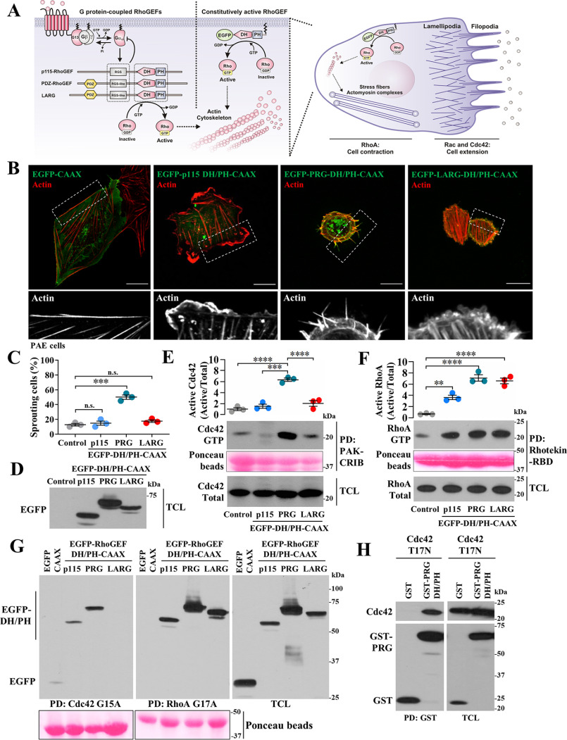
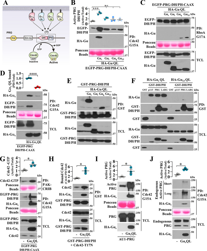
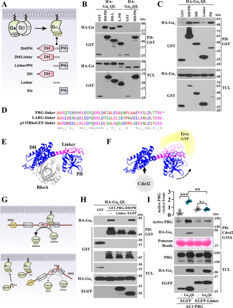
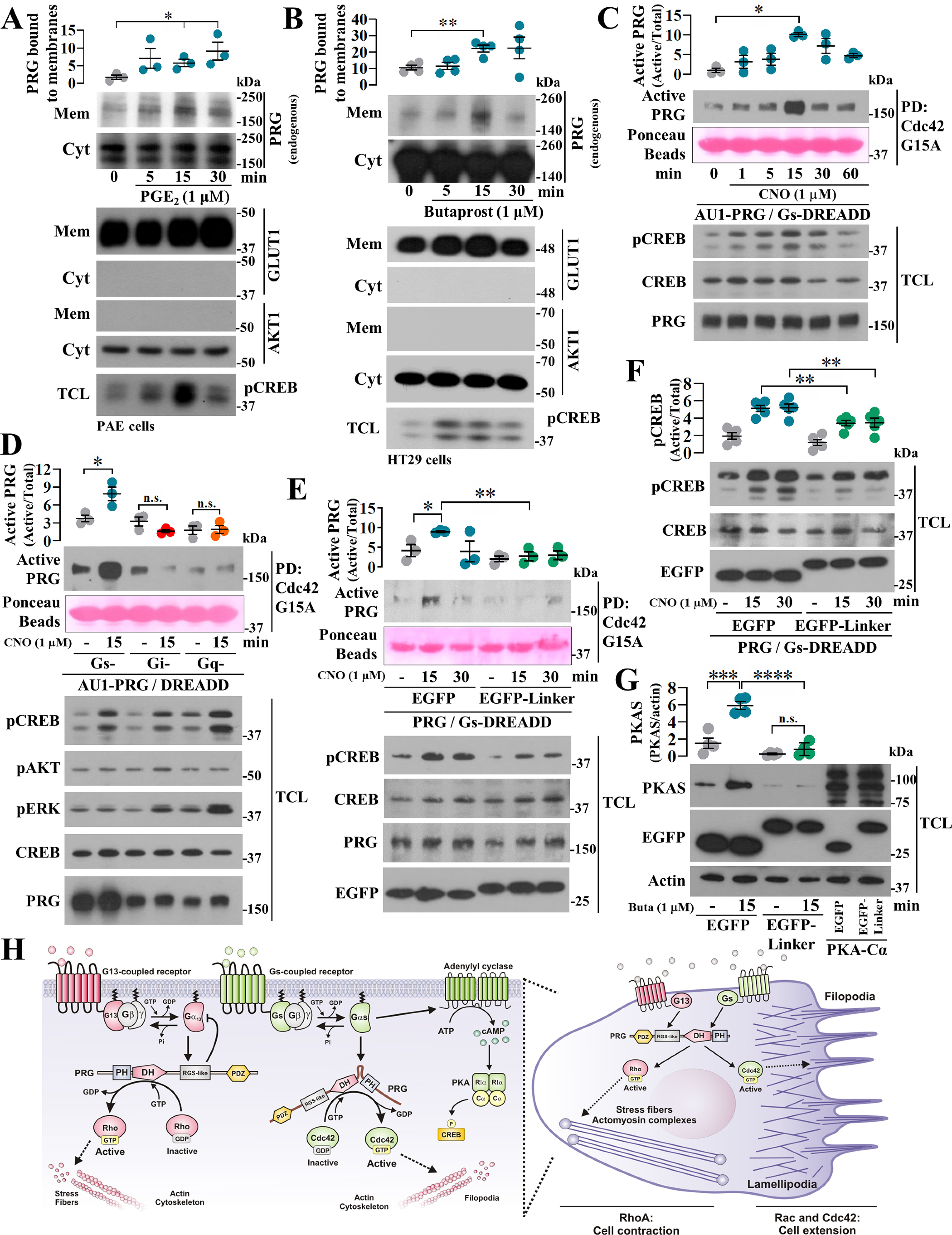
Gα proteins promote dynamic adjustments of cell shape directed by actin-cytoskeleton reorganization via their respective RhoGEF effectors. For example, Gα13 binding to the RGS-homology (RH) domains of several RH-RhoGEFs allosterically activates these proteins, causing them to expose their catalytic Dbl-homology (DH)/pleckstrin-homology (PH) regions, which triggers downstream signals. However, whether additional Gα proteins might directly regulate the RH-RhoGEFs was not known. To explore this question, we first examined the morphological effects of expressing shortened RH-RhoGEF DH/PH constructs of p115RhoGEF/ARHGEF1, PDZ-RhoGEF (PRG)/ARHGEF11, and LARG/ARHGEF12. As expected, the three constructs promoted cell contraction and activated RhoA, known to be downstream of Gα13 Intriguingly, PRG DH/PH also induced filopodia-like cell protrusions and activated Cdc42. This pathway was stimulated by constitutively active Gαs (GαsQ227L), which enabled endogenous PRG to gain affinity for Cdc42. A chemogenetic approach revealed that signaling by Gs-coupled receptors, but not by those coupled to Gi or Gq, enabled PRG to bind Cdc42. This receptor-dependent effect, as well as CREB phosphorylation, was blocked by a construct derived from the PRG:Gαs-binding region, PRG-linker. Active Gαs interacted with isolated PRG DH and PH domains and their linker. In addition, this construct interfered with GαsQ227L's ability to guide PRG's interaction with Cdc42. Endogenous Gs-coupled prostaglandin receptors stimulated PRG binding to membrane fractions and activated signaling to PKA, and this canonical endogenous pathway was attenuated by PRG-linker. Altogether, our results demonstrate that active Gαs can recognize PRG as a novel effector directing its DH/PH catalytic module to gain affinity for Cdc42.

Keywords: ARHGEF11; Cdc42; DH/PH catalytic module; G protein–coupled receptor (GPCR); GPCR; Galpha-s; Gαs; PDZ-RhoGEF; PDZ-RhoGEF (PRG); Rho (Rho GTPase); Rho GTPases; Rho guanine nucleotide exchange factor (RhoGEF); cell signaling; guanine nucleotide exchange factor (GEF); heterotrimeric G protein.


Conflict of interest statement

Conflict of interest—The authors declare that they have no conflicts of interest with the contents of this article.

Figures

Figure 1.
Figure 1.
In addition to its canonical effect on RhoA, PRG DH/PH activates Cdc42 and promotes filopodia formation. A, hypothetical effects of membrane-anchored RH-RhoGEF DH/PH catalytic modules. B, confocal images of PAE cells showing EGFP-RH-RhoGEF DH/PH-CAAX constructs (green) and their effects on F-actin (red). Zoomed-in areas are shown in the bottom row. Scale bar, 20 μm. C, graph shows the percentage of cells exhibiting filopodia-like structures. Each dot represents the means ± S.E. of at least 30 cells per experiment (n = 3). ***, p < 0.0001; n.s., no significance, one-way ANOVA followed Tukey. D, representative blot shows expression of EGFP-RH-RhoGEF DH/PH-CAAX constructs. E and F, activation of Cdc42 (E) and RhoA (F) by EGFP-RhoGEF DH/PH-CAAX constructs transfected into HEK293T cells was detected by PAK-CRIB and Rhotekin-RBD pulldown (PD), respectively. The graphs represent the means ± S.E. densitometric values (n = 3). **, p = 0.005; ***, p < 0.001; ****, p < 0.0001, one-way ANOVA followed Tukey. G, pulldown of active EGFP-RhoGEF DH/PH-CAAX constructs based on their affinity for nucleotide-free recombinant Cdc42-G15A (left panel) and RhoA-G17A (middle panel). Total cell lysates (TCL) are shown in the right panel. H, interaction between PRG DH/PH and Cdc42-T17N was assayed in transfected HEK293T cells subjected to pulldown assays (PD:GST). D–H, protein expression is confirmed in total cell lysates.
Figure 2.
Figure 2.
s-Q227L binds PRG DH/PH enabling this prototypical RhoA-specific GEF to directly activate Cdc42. A, hypothetic model postulating Gα subunits as potential regulators of PRG DH/PH catalytic module. B and C, the effect of GTPase-deficient Gα subunits on the interaction of EGFP-PRG-DH/PH-CAAX with Cdc42-G15A (B) and RhoA-G17A (C) was analyzed by pulldown (PD) using lysates from HEK293T cells transfected with HA-tagged Gαs, Gαi, Gαq, or Gα13 QL mutants and EGFP-PRG-DH/PH-CAAX. The graph in B represents the means ± S.E. (n = 3). ***, p < 0.001; n.s., no significance, one-way ANOVA followed Tukey. D, to address whether Gαs-QL detected in the PRG-DH/PH·Cdc42-G15A pulldown was part of a ternary complex, pulldown experiments were done in the presence or absence of PRG-DH/PH. The graph represents the means ± S.E. (n = 3). **, p < 0.0001, t test. E, the potential interaction between active Gα subunits and PRG DH/PH was analyzed in HEK293T cells transfected with GST-PRG-DH/PH and HA-tagged GTPase-deficient Gα subunits subjected to pulldown assays. F, interaction between Gαs-QL and the catalytic domain of the three RH-RhoGEFs was assayed by pulldown using HEK293T cells transfected with HA-Gαs-QL and GST-p115-DH/PH, GST-PRG-DH/PH, or GST-LARG-DH/PH. GST and HA-Gα13-QL served as negative controls. G, the effect of Gαs-QL on the activation of Cdc42 by PRG-DH/PH was assessed by pulldown using lysates of transfected HEK293T cells. The graph represents the means ± S.E. (n = 3). **, p = 0.01, t test. Representative blots show the fraction of active Cdc42 (top panel) and the active fraction of PRG-DH/PH with affinity for Cdc42-G15A (middle panel). H, the effect of Gαs-QL on the interaction between PRG-DH/PH and Cdc42-T17N was analyzed by pulldown using lysates of transfected HEK293T cells. The graph represents the means ± S.E. (n = 3). *, p = 0.01, t test. I and J, the effect of Gαs-QL on full-length PRG affinity for Cdc42 was analyzed in HEK293T cells that were transfected with full-length AU1-PRG (I) without or with HA-Gαs-QL or only with HA-Gαs-QL to address its effect on endogenous PRG (J). The active fraction of full-length PRG with affinity for Cdc42-G15A was isolated by pulldown and revealed by immunoblotting with anti-PRG antibodies. The graphs represents the means ± S.E. (n = 3). *, p = 0.01 in H and 0.04 in I, t test.
Figure 3.
Figure 3.
s-Q227L binds PRG DH and PH domains and the linker region joining them. A, model showing GST-tagged PRG-DH/PH constructs used to map Gαs·PRG-DH/PH interaction. B and C, interaction between HA-Gαs-QL and the indicated GST-PRG-DH/PH constructs was analyzed by pulldown using lysates of transfected HEK293T cells. HA-Gαs-QL and HA-Gα13-QL (used as control) were revealed with anti-HA antibodies. D, multiple alignment of p115RhoGEF, LARG, and PRG-linker regions. E, structure of PRG-DH/PH·RhoA complex (24). F, model showing Gαs-GTP·PRG-DH/PH complex; hypothetically, active Gαs constrains PRGDH/PH to bind Cdc42. G, model showing the potential inhibitory effect of the EGFP–PRG-linker construct on PRG activation by Gαs-QL. H, HEK293T cells transfected with EGFP-tagged PRG-linker construct (or EGFP) together with HA-Gαs-QL and GST-PRG-DH/PH (or GST) were subjected to GST pulldown assays. I, Gαs-dependent PRG·Cdc42 interaction was analyzed in HEK293T cells transfected with HA-Gαs-QL (or control plasmid) and AU1-PRG together with EGFP–PRG-linker or EGFP and subjected to Cdc42-G15A pulldown. The graph represents the means ± S.E. (n = 3). **, p = 0.001; ***, p = 0.0001; ns, no significance, one-way ANOVA followed Tukey.
Figure 4.
Figure 4.
Agonist-dependent stimulation of Gs-coupled receptors enables PRG to bind Cdc42. A and B, membrane recruitment of endogenous PRG promoted by Gs-coupled GPCR signaling was assessed in PAE (A) and HT29 (B) cells stimulated with 1 μm PGE2 or butaprost, respectively. PRG in membrane fractions was revealed by Western blotting. GLUT1 and AKT1 were used as membrane and cytosolic markers, respectively. The graphs represent the means ± S.E. (n = 3, *, p < 0.05 in A; and n = 4, **, p < 0.01 in B; t test). C, time course of PRG·Cdc42 interaction was assessed in COS7 expressing Gs-DREADD receptors. The cells were stimulated with 1 µm CNO and subjected to Cdc42-G15A pulldown. The graph represents the means ± S.E. (n = 3). *, p < 0.05, one-way ANOVA followed Tukey. D, the effect of different endogenous heterotrimeric G proteins on PRG affinity for Cdc42 was studied in COS7 cells transfected with AU1-PRG and Gs-, Gi-, or Gq-DREADDs. The cells were stimulated with CNO for 15 min and subjected to Cdc42-G15A pulldown assays. The graph represents the means ± S.E. (n = 3). *, p < 0.05, one-way ANOVA followed Tukey. E, effect of the PRG-linker construct on agonist-stimulated interaction between PRG and Cdc42 was assessed in COS7 cells transfected with Gs-DREADD, AU1-PRG, and EGFP–PRG-linker or EGFP. The cells were stimulated with CNO for 15 and 30 min and subjected to Cdc42-G15A pulldown. The graph represents the means ± S.E. (n = 3). *, p = 0.0342; **, p = 0.0056, t test. F, effect of PRG-linker on agonist-dependent phosphorylation of CREB was assessed in COS7 cells expressing Gs-DREADDs and stimulated with CNO. The graph represents the means ± S.E. (n = 5). **, p < 0.01, t test. G, agonist-dependent phosphorylation of PKA substrates was assessed using PAE cells expressing EGFP or EGFP–PRG-linker and stimulated with butaprost. Lysates from EGFP-PKA-Cα−transfected cells served as control to detect PKA substrates. The graph represents the means ± S.E. (n = 4). ***, p = 0.0009; ****, p < 0.0001; n.s., no significance, one-way ANOVA followed Tukey. H, model depicts the canonical G13-PRG signaling axis to Rho and the emerging GPCR–Gαs–PRG–Cdc42 pathway based on the current findings. In cells, both systems putatively guide dynamic adjustments on actin-cytoskeleton reorganization.

Comment in

Similar articles

Cited by

References

    1. Ridley, A. J., Schwartz, M. A., Burridge, K., Firtel, R. A., Ginsberg, M. H., Borisy, G., Parsons, J. T., and Horwitz, A. R. (2003) Cell migration: integrating signals from front to back. Science 302, 1704–1709 10.1126/science.1092053 - DOI - PubMed
    1. Pollard, T. D., and Borisy, G. G. (2003) Cellular motility driven by assembly and disassembly of actin filaments. Cell 112, 453–465 10.1016/S0092-8674(03)00120-X - DOI - PubMed
    1. Aittaleb, M., Boguth, C. A., and Tesmer, J. J. (2010) Structure and function of heterotrimeric G protein-regulated Rho guanine nucleotide exchange factors. Mol. Pharmacol. 77, 111–125 10.1124/mol.109.061234 - DOI - PMC - PubMed
    1. Cook, D. R., Rossman, K. L., and Der, C. J. (2014) Rho guanine nucleotide exchange factors: regulators of Rho GTPase activity in development and disease. Oncogene 33, 4021–4035 10.1038/onc.2013.362 - DOI - PMC - PubMed
    1. Lawson, C. D., and Ridley, A. J. (2018) Rho GTPase signaling complexes in cell migration and invasion. J. Cell Biol. 217, 447–457 10.1083/jcb.201612069 - DOI - PMC - PubMed

Publication types

Substances