Review
. 2020 Oct;216(2):269-313.
doi: 10.1534/genetics.120.303095.

Regulation of Body Size and Growth Control

Affiliations
Review

Regulation of Body Size and Growth Control

Michael J Texada et al. Genetics. 2020 Oct.

Abstract

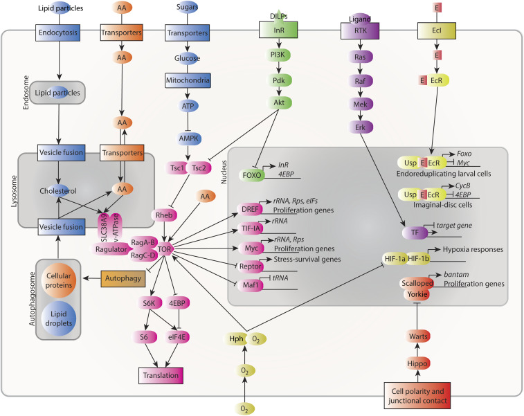
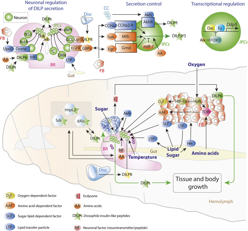
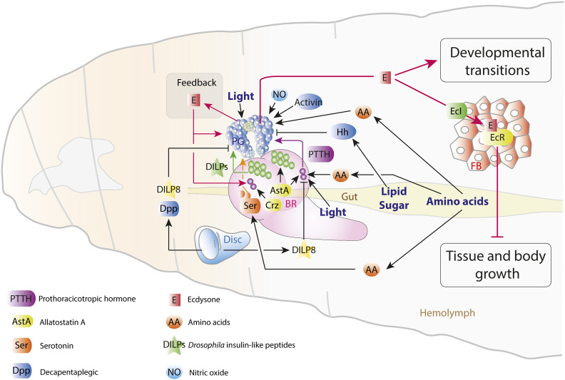
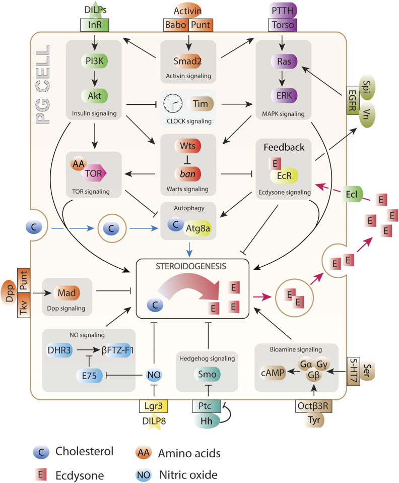
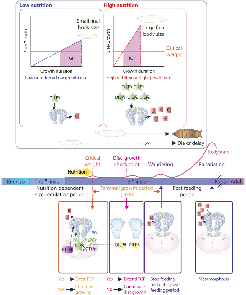
The control of body and organ growth is essential for the development of adults with proper size and proportions, which is important for survival and reproduction. In animals, adult body size is determined by the rate and duration of juvenile growth, which are influenced by the environment. In nutrient-scarce environments in which more time is needed for growth, the juvenile growth period can be extended by delaying maturation, whereas juvenile development is rapidly completed in nutrient-rich conditions. This flexibility requires the integration of environmental cues with developmental signals that govern internal checkpoints to ensure that maturation does not begin until sufficient tissue growth has occurred to reach a proper adult size. The Target of Rapamycin (TOR) pathway is the primary cell-autonomous nutrient sensor, while circulating hormones such as steroids and insulin-like growth factors are the main systemic regulators of growth and maturation in animals. We discuss recent findings in Drosophila melanogaster showing that cell-autonomous environment and growth-sensing mechanisms, involving TOR and other growth-regulatory pathways, that converge on insulin and steroid relay centers are responsible for adjusting systemic growth, and development, in response to external and internal conditions. In addition to this, proper organ growth is also monitored and coordinated with whole-body growth and the timing of maturation through modulation of steroid signaling. This coordination involves interorgan communication mediated by Drosophila insulin-like peptide 8 in response to tissue growth status. Together, these multiple nutritional and developmental cues feed into neuroendocrine hubs controlling insulin and steroid signaling, serving as checkpoints at which developmental progression toward maturation can be delayed. This review focuses on these mechanisms by which external and internal conditions can modulate developmental growth and ensure proper adult body size, and highlights the conserved architecture of this system, which has made Drosophila a prime model for understanding the coordination of growth and maturation in animals.

Keywords: DILP8; Drosophila; FlyBook; PTTH; checkpoint; critical weight; ecdysone; insulin; metamorphosis; prothoracic gland; timing.


Figures

Figure 1
Figure 1
The development of D. melanogaster. A fertilized Drosophila embryo spends roughly 1 day developing into a mobile, feeding larva (under normal conditions). After hatching, the larva feeds for the next 4 days, growing to 200 times its initial size; to accommodate this dramatic growth, the larva sheds its cuticle twice during this time in molts that separate the first, second, and third larval “instars.” After larval growth is complete, the animal wanders away from its food source to find a location suitable for the 4-day metamorphosis period, during which time the animal survives on stored material while its larval tissues are degraded and adult structures finish their development. The adult emerges (“ecloses”) once this process is complete. Pulses of the insect steroid hormone ecdysone regulate the animal’s progression through these developmental stages.
Figure 2
Figure 2
Intracellular signaling pathways govern cell growth and proliferation. Cholesterol (blue), amino acids (orange), sugars (blue), and oxygen (olive) feed into growth regulation through cell-autonomous regulation of TOR signaling (pink; some aspects of the TOR pathway in this diagram are mammal-specific, such as SLC38A9-mediated regulation; there is no close Drosophila ortholog of this protein). Local signaling via the Hippo/Warts pathway (reddish orange) responds to cell–cell junctions and epithelial organization, and receptor tyrosine kinase signaling (purple) responds to systemic or local signals. Systemic signaling through insulin-like factors (green) and ecdysone (E, yellow) also governs cell activity. Pathways are shown terminating in the nucleus with transcription factor activity. Not all pathway components are shown, and most links between pathways are not shown. eIFs, eukaryotic initiation factors; Rps, ribosomal proteins; rRNA, ribosomal RNA; tRNA, transfer RNA.
Figure 3
Figure 3
Regulation of insulin expression, release, and activity in the Drosophila larva. The larval insulin-producing cells (IPCs, small green spheres) of the brain (pink) receive a multitude of regulatory inputs (see also Table 1). Bottom panel: signals released by the fat body (FB), the gut, the developing imaginal tissues, and the prothoracic gland (PG) act on the IPCs to regulate DILP expression and release. Top left panel: input from neurons that sense temperature, disc development, and humoral factors act on the IPCs. BR, brain; DRN, DILP2-recruiting neurons; GCL, growth-coordinating Lgr3+ neurons. Top middle panel: Akh/AkhR signaling in the larval IPCs promotes DILP3 release; DILP2 and DILP5 are regulated by fat-derived activating factors CCHa2 and Stunted, which signal “nutrition,” and the inhibitor Eiger, which conveys “starvation.” Top, right panel: little is known about the cis-regulation of insulin gene expression. Dachshund and Eyeless, like their mammalian homologs Dach1/2 and Pax6, promote insulin expression, specifically of Dilp5. This expression is inhibited by FOXO, and signaling through the receptor Alk derepresses Dilp5 in response to the ligand Jelly Belly released by glia of the blood/brain barrier during starvation.
Figure 4
Figure 4
Regulation and effects of ecdysone (E) production in Drosophila larvae. A network of signals regulates E production in the prothoracic gland (PG). Nutritional influences (relayed by Hh, AstA, Crz, and amino acid-regulated serotonergic neurons) act on the IPCs, the PTTHn (prothoracicotropic hormone-producing neurons), and the PG; signals from the developing imaginal discs (DILP8 and Dpp) act on these cells as part of the growth-coordination mechanism. Light and internal clocks (not shown) regulate the PG and the PTTHn. E feeds back onto the PG to upregulate and then downregulate its own production, and onto the PTTHn to promote PTTH expression. Peaks of E act to promote developmental transitions, and basal levels block the growth of larval tissues while promoting disc growth. E entry is mediated by the E importer, EcI. See also Table 2 and Table 3.
Figure 5
Figure 5
Pathways affecting ecdysone (E) synthesis and release in the Drosophila prothoracic gland (PG). A broad array of autonomous and external cues govern the production and release of E, both at basal levels that regulate the growth of larval and imaginal tissues as well as in the peaks of synthesis that govern developmental transitions. DILP and PTTH signals carry nutritional and developmental information; the competence of the PG to respond to these signals is regulated by Activin signaling. Nutrition also affects the PG through the TOR and Warts pathways, as well as through inputs from serotonergic neurons and gut-derived Hedgehog (Hh). The metabolic state of the PG regulates cholesterol trafficking for steroidogenesis, and the developmental state of imaginal tissues is conveyed directly to the PG by the disc-derived factors DILP8 and Dpp (as well as by indirect means such as PTTH). PG-autonomous molecular clocks interface with external clock input (not shown) to organize E pulses. Feedback through E (via Ecdysone Importer, EcI) and EGF-like ligands drives and sculpts E peaks. See also Table 3.
Figure 6
Figure 6
Developmental checkpoints determine developmental timing in Drosophila. Top: at the onset of the final (third) instar, the resumption of larval feeding after the previous molt stimulates a small rise in ecdysone (E) production via insulin together with PTTH. This small ecdysone pulse results in attainment of the nutrition-sensitive developmental checkpoint critical weight (CW), which begins the subsequent nonnutrition-sensitive feeding period called the terminal growth period (TGP). In post-CW larvae, further nutrition intake is not necessary to undergo metamorphosis. Larvae will not proceed into metamorphosis until they reach CW, which reflects their ability to survive through pupal life on stored nutrients alone. After CW, the larva continues to grow during TGP if food is present. Under poor conditions (left), the larva grows slowly, reaching CW later; slow growth continues during TGP, leading to small adults. This slow growth and delayed maturation results from low production of insulin and ecdysone. Right: under rich conditions, the larva feeds well and produces more insulin, which induces fast growth. The animal soon reaches CW and continues to grow quickly during TGP. Insulin also promotes earlier peaks of ecdysone, which accelerate developmental timing. As a result, adults emerge more quickly, with larger bodies. Bottom: organ growth also affects developmental timing. Developing discs secrete DILP8, which affects the timing of PTTH secretion and indirectly controls the timing of growth cessation by regulating the timing of the second small ecdysone pulse. DILP8 also regulates the growth of all discs simultaneously by acting directly on the prothoracic gland (PG) to regulate basal ecdysone levels.

Similar articles

Cited by

References

    1. Acevedo-Rodriguez A., Kauffman A. S., Cherrington B. D., Borges C. S., Roepke T. A. et al. , 2018. Emerging insights into hypothalamic-pituitary-gonadal axis regulation and interaction with stress signalling. J. Neuroendocrinol. 30: e12590 10.1111/jne.12590 - DOI - PMC - PubMed
    1. Agbu P., Cassidy J. J., Braverman J., Jacobson A., and Carthew R. W., 2020. MicroRNA miR-7 regulates secretion of insulin-like peptides. Endocrinology 161: bqz040. 10.1210/endocr/bqz040 - DOI - PMC - PubMed
    1. Agrawal N., Delanoue R., Mauri A., Basco D., Pasco M. et al. , 2016. The Drosophila TNF Eiger is an adipokine that acts on insulin-producing cells to mediate nutrient response. Cell Metab. 23: 675–684. 10.1016/j.cmet.2016.03.003 - DOI - PubMed
    1. Ahmed M. L., Ong K. K., and Dunger D. B., 2009. Childhood obesity and the timing of puberty. Trends Endocrinol. Metab. 20: 237–242. 10.1016/j.tem.2009.02.004 - DOI - PubMed
    1. Ajuria L., Nieva C., Winkler C., Kuo D., Samper N. et al. , 2011. Capicua DNA-binding sites are general response elements for RTK signaling in Drosophila. Development 138: 915–924. 10.1242/dev.057729 - DOI - PMC - PubMed

Publication types

MeSH terms

LinkOut - more resources