Suppression of pancreatic cancer liver metastasis by secretion-deficient ITIH5
- PMID: 33024269
- PMCID: PMC7782545
- DOI: 10.1038/s41416-020-01093-z
Suppression of pancreatic cancer liver metastasis by secretion-deficient ITIH5
Abstract
Background: Previously, we identified ITIH5 as a suppressor of pancreatic ductal adenocarcinoma (PDAC) metastasis in experimental models. Expression of ITIH5 correlated with decreased cell motility, invasion and metastasis without significant inhibition of primary tumour growth. Here, we tested whether secretion of ITIH5 is required to suppress liver metastasis and sought to understand the role of ITIH5 in human PDAC.
Methods: We expressed mutant ITIH5 with deletion of the N-terminal secretion sequence (ITIH5Δs) in highly metastatic human PDAC cell lines. We used a human tissue microarray (TMA) to compare ITIH5 levels in uninvolved pancreas, primary and metastatic PDAC.
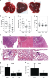
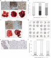
Results: Secretion-deficient ITIH5Δs was sufficient to suppress liver metastasis. Similar to secreted ITIH5, expression of ITIH5Δs was associated with rounded cell morphology, reduced cell motility and reduction of liver metastasis. Expression of ITIH5 is low in both human primary PDAC and matched metastases.
Conclusions: Metastasis suppression by ITIH5 may be mediated by an intracellular mechanism. In human PDAC, loss of ITIH5 may be an early event and ITIH5-low PDAC cells in primary tumours may be selected for liver metastasis. Further defining the ITIH5-mediated pathway in PDAC could establish future therapeutic exploitation of this biology and reduce morbidity and mortality associated with PDAC metastasis.
Conflict of interest statement
The authors declare no competing interests.
Figures


Similar articles
-
An Overview on the Emerging Role of the Plasma Protease Inhibitor Protein ITIH5 as a Metastasis Suppressor.Cell Biochem Biophys. 2024 Jun;82(2):399-409. doi: 10.1007/s12013-024-01227-7. Epub 2024 Feb 15. Cell Biochem Biophys. 2024. PMID: 38355846 Review.
-
Genome-wide in vivo RNAi screen identifies ITIH5 as a metastasis suppressor in pancreatic cancer.Clin Exp Metastasis. 2017 Apr;34(3-4):229-239. doi: 10.1007/s10585-017-9840-3. Epub 2017 Mar 13. Clin Exp Metastasis. 2017. PMID: 28289921 Free PMC article.
-
Small Nucleolar Noncoding RNA SNORA23, Up-Regulated in Human Pancreatic Ductal Adenocarcinoma, Regulates Expression of Spectrin Repeat-Containing Nuclear Envelope 2 to Promote Growth and Metastasis of Xenograft Tumors in Mice.Gastroenterology. 2017 Jul;153(1):292-306.e2. doi: 10.1053/j.gastro.2017.03.050. Epub 2017 Apr 5. Gastroenterology. 2017. PMID: 28390868
-
The Novel KLF4/MSI2 Signaling Pathway Regulates Growth and Metastasis of Pancreatic Cancer.Clin Cancer Res. 2017 Feb 1;23(3):687-696. doi: 10.1158/1078-0432.CCR-16-1064. Epub 2016 Jul 22. Clin Cancer Res. 2017. PMID: 27449499 Free PMC article.
-
UBL4A inhibits autophagy-mediated proliferation and metastasis of pancreatic ductal adenocarcinoma via targeting LAMP1.J Exp Clin Cancer Res. 2019 Jul 9;38(1):297. doi: 10.1186/s13046-019-1278-9. J Exp Clin Cancer Res. 2019. PMID: 31288830 Free PMC article.
Cited by
-
ITIH5 as a multifaceted player in pancreatic cancer suppression, impairing tyrosine kinase signaling, cell adhesion and migration.Mol Oncol. 2024 Jun;18(6):1486-1509. doi: 10.1002/1878-0261.13609. Epub 2024 Feb 20. Mol Oncol. 2024. PMID: 38375974 Free PMC article.
-
ITIH5, a p53-responsive gene, inhibits the growth and metastasis of melanoma cells by downregulating the transcriptional activity of KLF4.Cell Death Dis. 2021 May 2;12(5):438. doi: 10.1038/s41419-021-03707-7. Cell Death Dis. 2021. PMID: 33935281 Free PMC article.
-
Serum ITIH5 as a novel diagnostic biomarker in cholangiocarcinoma.Cancer Sci. 2024 May;115(5):1665-1679. doi: 10.1111/cas.16143. Epub 2024 Mar 12. Cancer Sci. 2024. PMID: 38475675 Free PMC article.
-
An Overview on the Emerging Role of the Plasma Protease Inhibitor Protein ITIH5 as a Metastasis Suppressor.Cell Biochem Biophys. 2024 Jun;82(2):399-409. doi: 10.1007/s12013-024-01227-7. Epub 2024 Feb 15. Cell Biochem Biophys. 2024. PMID: 38355846 Review.
-
Risk factors and predictive nomograms for early death of patients with pancreatic cancer liver metastasis: A large cohort study based on the SEER database and Chinese population.Front Oncol. 2022 Sep 23;12:998445. doi: 10.3389/fonc.2022.998445. eCollection 2022. Front Oncol. 2022. PMID: 36212438 Free PMC article.
References
Publication types
MeSH terms
Substances
Grants and funding
LinkOut - more resources
Full Text Sources
Medical
Molecular Biology Databases

