Review
. 2020 Jul 27;36(5):837-844.
doi: 10.1002/joa3.12410. eCollection 2020 Oct.

Junctional ectopic tachycardia (JET)

Affiliations
Review

Junctional ectopic tachycardia (JET)

Mohammad Alasti et al. J Arrhythm. .

Abstract

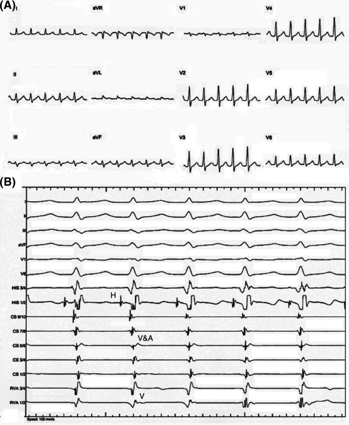
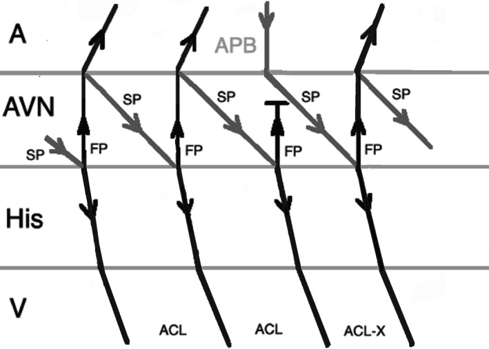
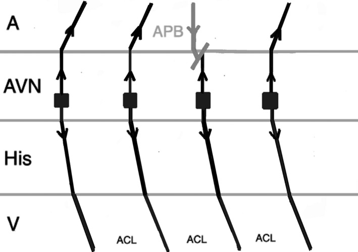
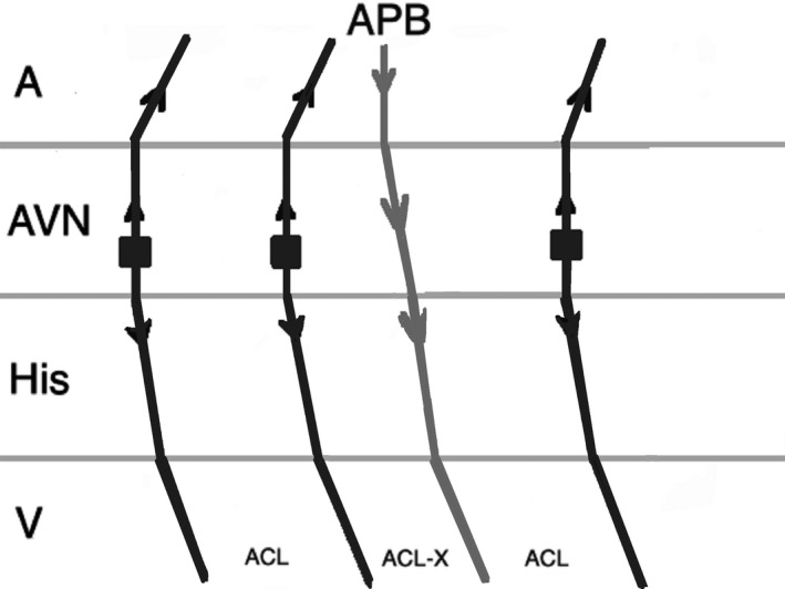
Junctional ectopic tachycardia (JET) is a tachyarrhythmia arising from the atrioventricular node and His bundle area. Enhanced normal automaticity has been postulated as the mechanism of JET in the majority of patients. It is more common in children and can be seen as congenital or in postoperative settings. It is often a narrow complex tachycardia but can present as a wide complex tachycardia as a result of aberrant conduction. Its differentiation from other arrhythmias especially atrioventricular nodal reentrant tachycardia (AVNRT) can be challenging. Medical treatment of JET is difficult, and catheter ablation remains the mainstay of treatment in refractory cases with a high risk of atrioventricular block and recurrence.

Keywords: arrhythmia; ectopic; focal; junctional tachycardia; pediatrics; postoperative.


Conflict of interest statement

The authors declare no conflict of interests for this article.

Figures

FIGURE 1
FIGURE 1
A, Twelve‐lead ECG showing a regular narrow complex tachycardia with a cycle length of 412 milliseconds and no obvious P waves before QRS complexes. B, Intracardiac recordings showing a tachycardia with one‐to‐one VA association and a cycle length of 412 milliseconds, HV interval of 45 milliseconds, and very short VA interval (A, atrial; H, His; V, ventricular)
FIGURE 2
FIGURE 2
Response to an atrial premature beat delivered during His refractoriness (late APB) in AVNRT which does not influence the immediate beat but advances the next beat through conduction via slow pathway. (A, atrium; ACL, arrhythmia cycle length; APB, atrial premature beat; AVN, atrioventricular node; FP, fast pathway; SP, slow pathway; V, ventricle)
FIGURE 3
FIGURE 3
Response to a late APB in JET which does not influence the immediate and the next beats. (A, atrium; ACL, arrhythmia cycle length; APB, atrial premature beat; AVN, atrioventricular node; V, ventricle)
FIGURE 4
FIGURE 4
Response to an atrial premature beat delivered before His refractoriness (early APB) in JET which influences the immediate beat through conduction via fast pathway and the arrhythmia continues. (A, atrium; ACL, arrhythmia cycle length; APB, atrial premature beat; AVN, atrioventricular node; V, ventricle)
FIGURE 5
FIGURE 5
Response to an early APB in AVNRT which influences the immediate His through conduction via fast pathway and the arrhythmia terminates as a result of fast pathway refractoriness. (A, atrium; ACL, arrhythmia cycle length; APB, atrial premature beat; AVN, atrioventricular node; FP, fast pathway; SP, slow pathway; V, ventricle)
FIGURE 6
FIGURE 6
Response to an early APB in AVNRT with simultaneous anterograde fast and slow AV nodal pathways conduction. Therefore, AVNRT continues after an APB that advances the immediate His. (A, atrium; ACL, arrhythmia cycle length; APB, atrial premature beat; AVN, atrioventricular node; FP, fast pathway; SP, slow pathway; V, ventricle)
FIGURE 7
FIGURE 7
Response to an atrial premature beat delivered during His refractoriness in JET when dual AV node physiology is present. It can lead to misdiagnosis of JET as AVNRT. (A, atrium; ACL, arrhythmia cycle length; APB, atrial premature beat; AVN, atrioventricular node; FP, fast pathway; RCL, return cycle length; SP, slow pathway; V, ventricle)
FIGURE 8
FIGURE 8
Response to atrial overdrive pacing in JET: after termination of pacing, the first return beat (arrhythmia) begins with an H followed by an A (A‐H‐H‐A response). (A, atrium; ACL, arrhythmia cycle length; AVN, atrioventricular node; FP, fast pathway; H, His; PCL, pacing cycle length; RCL, return cycle length; V, ventricle)
FIGURE 9
FIGURE 9
Response to atrial overdrive pacing in AVNRT: after termination of pacing, the first return beat begins with an A (A‐H‐A response). (A, atrium; ACL, arrhythmia cycle length; AVN, atrioventricular node; FP, fast pathway; H, His, PCL, pacing cycle length, RCL, return cycle length, SP, slow pathway, V, ventricle)
FIGURE 10
FIGURE 10
A pseudo A‐H‐H‐A response after overdrive pacing in AVNRT. The first beat after stopping pacing is not related to the last atrial paced beat as a result of slow conduction via slow pathway. (A, atrium; ACL, arrhythmia cycle length; AVN, atrioventricular node; FP, fast pathway; H, His; PCL, pacing cycle length; RCL, return cycle length; SP, slow pathway; V, ventricle)
FIGURE 11
FIGURE 11
An A‐H‐H‐A response after overdrive pacing in AVNRT. During atrial pacing, the tachycardia (arrhythmia 1) is terminated and the last atrial paced beat results in two His signals as conduction proceeds anterograde down both the fast pathway and slow pathway and reinitiates the tachycardia (arrhythmia 2). (A, atrium; ACL1, arrhythmia 1 cycle length; ACL2, arrhythmia 2 cycle length; AVN, atrioventricular node; FP, fast pathway; H, His; PCL, pacing cycle length; SP, slow pathway; V, ventricle)
FIGURE 12
FIGURE 12
An A‐H‐A response after overdrive pacing in JET. When anterograde fast pathway conduction is poor, atrial overdrive pacing during JET conducts down the slow pathway and up the fast pathway causing an echo beat, results in an A‐H‐A response. (A, atrium; ACL, arrhythmia cycle length; AVN, atrioventricular node; FP, fast pathway; H, His; PCL, pacing cycle length; RCL, return cycle length; SP, slow pathway; V, ventricle)

References

    1. Liu CF, Ip JE, Markowitz SM, Lerman BB. Junctional tachycardia In: Zipes D, Jalife J, Stevenson W, editors. Cardiac electrophysiology: from cell to bedside, 7th edn Philadelphia, PA: Elsevier, 2018; p. 768–775.
    1. Gonzalez MD, Banchs JE, Moukabary T, Rivera J. Ablation of atrioventricular junctional tachycardia: atrioventricular variants and focal junctional tachycardia In: Huang SKS, Miller JM, editors. Catheter ablation of cardiac arrhythmias, 4th edn Philadelphia, PA: Elsevier, 2020; p. 316–348.
    1. Collins KK, Van Hare GF, Kertesz NJ, Law IH, Bar‐Cohen Y, Dubin AM, Etheridge SP, Berul CI, Avari JN, Tuzcu V, Sreeram N, Schaffer MS, Fournier A, Sanatani S, Snyder CS, Smith RT, Arabia L, Hamilton R, Chun T, Liberman L, Kakavand B, Paul T, Tanel RE. Pediatric non post‐operative junctional ectopic tachycardia: Medical management and interventional therapies. J Am Coll Cardiol. 2009;53(8):690–7. - PubMed
    1. Ozyilmaz EY, Ozyilmaz S, Guzeltas A. Junctional ectopic tachycardia in late period after early postoperative complete atrioventricular block: Messenger of return to normal sinus rhythm? Explanation with four case series. J Electrocardiol. 2017;50:378–382. - PubMed
    1. Pierick AR, Law IH, Muldonado JR, Bergen NH. Junctional ectopic tachycardia localization and procedural approach using cryoablation. Pacing Clin Electrophysiol. 2017;40:655–660. - PubMed