. 2020 Sep 2;9(9):e1397-e1408.
doi: 10.1016/j.eats.2020.05.024. eCollection 2020 Sep.

Latarjet Cerclage: The Metal-Free Fixation

Affiliations

Latarjet Cerclage: The Metal-Free Fixation

Abdul-Ilah Hachem et al. Arthrosc Tech. .

Abstract

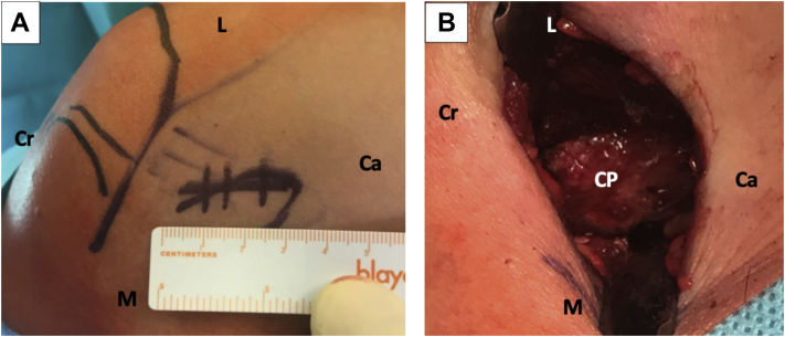
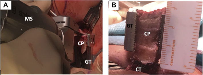
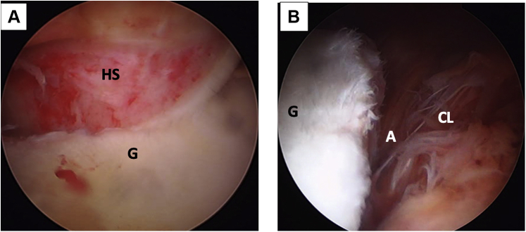
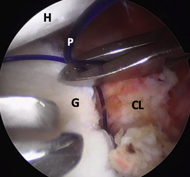
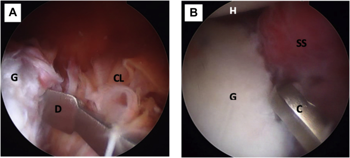
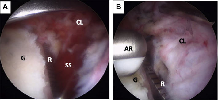
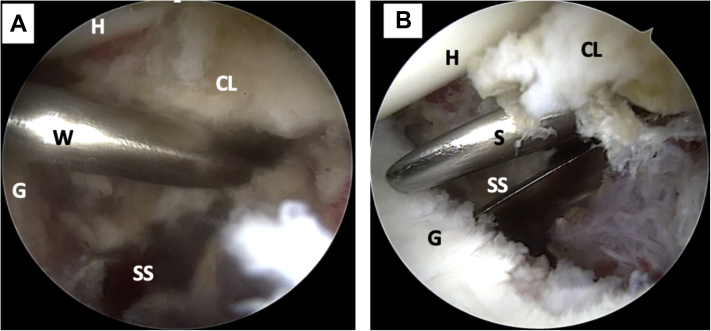
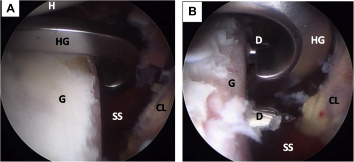
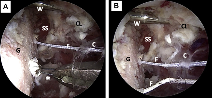
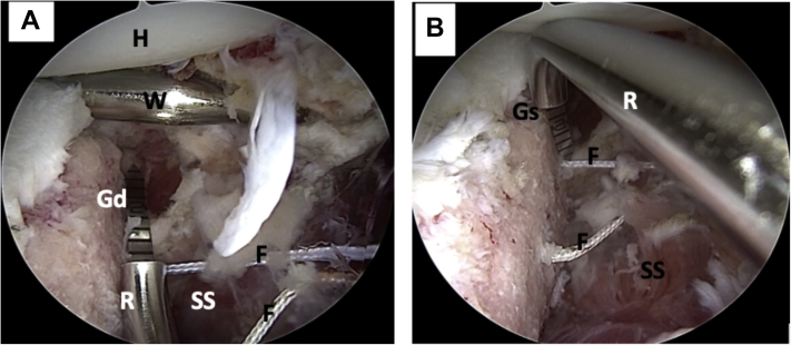
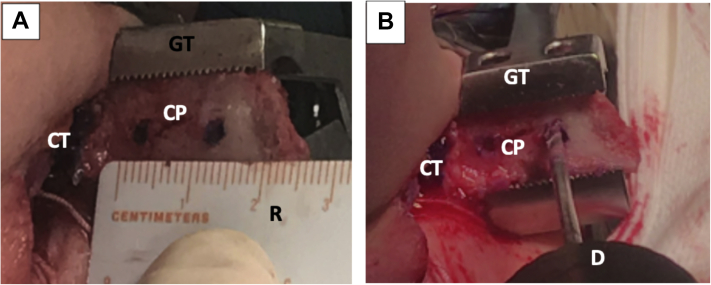
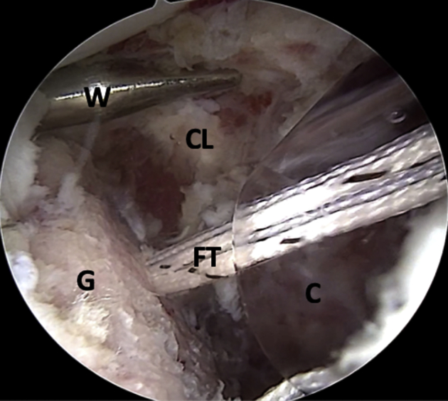
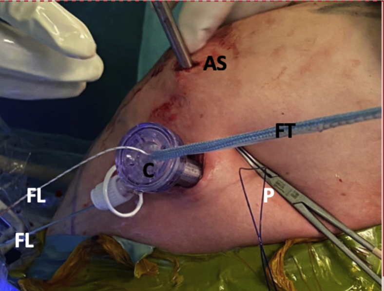
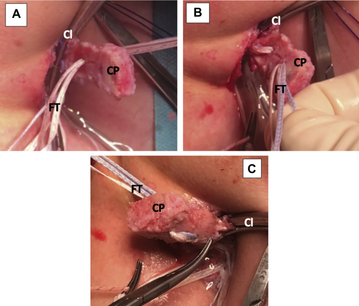
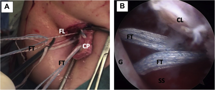
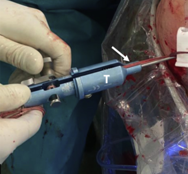
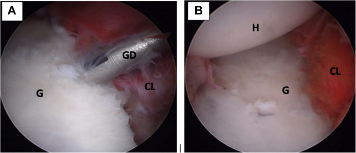
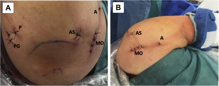
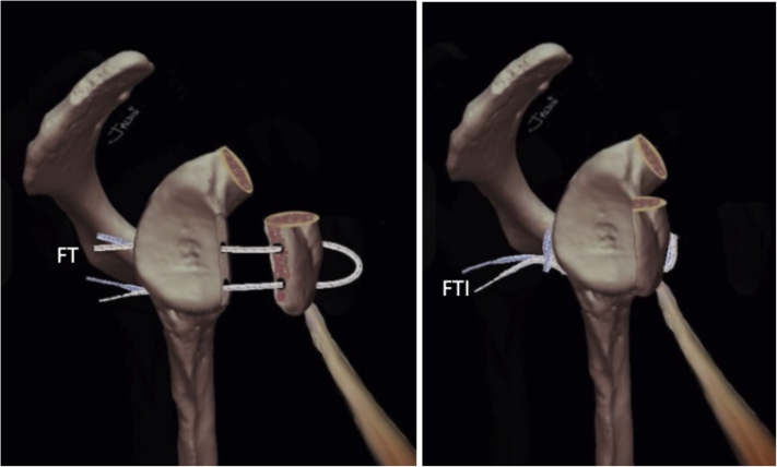
The Latarjet technique is a widely used technique for anterior shoulder instability with glenoid bone defects, irreparable capsuloligamentous lesion, or in patients at greater risk of recurrence. The use of this technique has been reported to obtain satisfactory clinical and biomechanical results. Although other methods exist, the coracoid process is typically fixed with 2 metal screws. Complications related to metal fixation are very frequently reported. In an attempt to avoid these complications, we developed this arthroscopically assisted metal-free Latarjet technique in which we fix a coracoid graft using four cerclage tapes to achieve a strong, stable fixation, thus mimicking a plate.


Figures

Fig 1
Fig 1
Left shoulder, lateral decubitus position, anterior view. (A) Direct mini-open skin incision of 3 cm over the coracoid process (not seen on image). (B) Mini-open approach; the coracoacromial ligament and the pectoralis minor muscle attachments are released from the coracoid process. (Ca, caudal; CP, coracoid process; Cr, cranial; L, lateral; M, medial.)
Fig 2
Fig 2
Left shoulder, lateral decubitus position, anterior view. (A) The Inferior part of the coracoid process is flattened with a motorized saw, while the graft is immobilized with a grabber tool. (B) Through the mini-open approach, the coracoid process is externalized and measured. Notice the conjoint tendon attached to the coracoid’s process base. (CP, coracoid process; CT, conjoint tendon; GT, grabber tool; MS, motorized saw.)
Fig 3
Fig 3
(A) Left shoulder, lateral decubitus position, posterior portal, posterolateral humeral head - Hill Sachs lesion engaging on anterior glenoid rim. (B) Arthroscopic view, anterosuperior portal. Anterior glenoid rim with an anterior labroligamentous periosteal sleeve avulsion lesion. (A, anterior labroligamentous periosteal sleeve avulsion lesion; CL, capsulolabral complex; G, anterior glenoid rim; HS, Hill–Sachs lesion.)
Fig 4
Fig 4
Left shoulder, lateral decubitus position, arthroscopic view, anterosuperior portal. A polydioxanone suture is passed through anterior capsulolabral complex to pull it anteriorly and obtain a wide view of the anterior glenoid rim. (CL, capsulolabral complex; G, anterior glenoid rim; H, humeral head; P, polydioxanone suture.)
Fig 5
Fig 5
Left shoulder, lateral decubitus position, arthroscopic view, anterosuperior portal. (A) The anterior capsulolabral complex is detached from the anterior glenoid rim with a dissector. (B) The anterior glenoid rim is prepared with a curette for biological healing of the re-attached capsulolabral complex. (C, curette; CL, capsulolabral complex; D, dissector; G, anterior glenoid rim; H, humeral head; SS, subscapularis muscle.)
Fig 6
Fig 6
Left shoulder, lateral decubitus position, arthroscopic view, anterosuperior portal. (A) The anterior glenoid defect is measured with a ruler starting from the lower edge of the defect. The anterior traction of the capsulolabral complex, done with a polydioxanone suture (not on image), can also be seen. (B) The anterior glenoid defect (G) is marked with an arthroscopic radiofrequency at the midpoint of the length of the coracoid process (not on image). (AR, arthroscopic radiofrequency; CL, capsulolabral complex; G, anterior glenoid rim; R, ruler; SS, subscapularis muscle.)
Fig 7
Fig 7
Left shoulder, lateral decubitus position, arthroscopic view, anterosuperior portal. (A) A Wissinger rod is passed from the posterior portal to mark the level of the subscapularis muscle split (lower part). (B) Arthroscopic view, anterosuperior portal. The subscapularis muscle split is made using scissors. (CL, capsulolabral complex; G, glenoid; H, humeral head; S, scissors; SS, subscapularis muscle; W, Wissinger rod.)
Fig 8
Fig 8
Left shoulder, lateral decubitus position, arthroscopic view, anterosuperior portal. (A) A metal hook guide is introduced posteriorly and positioned over the mark on the anterior glenoid rim. (B) Using the hook guide, 2 tunnels are made with 2 cannulated 2.4-mm drills. (CL, capsulolabral complex; D, drill; G, anterior glenoid rim; H, humeral head; HG, metal hook guide; SS, subscapularis muscle.)
Fig 9
Fig 9
Left shoulder, lateral decubitus position, arthroscopic view, anterosuperior portal. (A) The cannulated drills have been removed; nitinol wires are used to transport the FiberLink sutures. The Wissinger rod marking the level of the subscapularis split where the cannula is positioned can be noted. (B) Nitinol wires are replaced with FiberLink sutures. The FiberLink sutures are of different colors to avoid mistakes in handling. ∗Nitinol wire (C, cannula; CL, capsulolabral complex; F, FiberLink sutures; G, glenoid; SS, subscapularis muscle; W, wissinger rod.)
Fig 10
Fig 10
Left shoulder, lateral decubitus position, arthroscopic view, anterosuperior portal. (A) The distance from the inferior tunnel to the most distal limit of the anterior glenoid defect is measured. (B) The distance from the glenoid surface to the tunnels is measured with a ruler. (F, FiberLink sutures; Gd, glenoid defect; Gs, glenoid surface; H, humeral head; R, ruler; SS, subscapularis muscle; W, Wissinger rod.)
Fig 11
Fig 11
Left shoulder, lateral decubitus position, anterior view. Externalized coracoid process through mini-open approach. (A) The positions of the tunnels are marked on the coracoid process while it is manipulated with a grabber tool. (B) Two tunnels are drilled over the marks. (CP, coracoid process; CT, conjoint tendon; D, drill; GT, grabber tool; R, ruler.)
Fig 12
Fig 12
Left shoulder, lateral decubitus position, arthroscopic view, anterosuperior portal. The FiberLink sutures (not on image) are pulled through the cannula to transport the FiberTape cerclage tapes. (C, cannula; CL, capsulolabral complex; FT, FiberTape cerclage tapes; G, glenoid; W, Wissinger rod.)
Fig 13
Fig 13
Left shoulder, lateral decubitus position, anterior view. An 8.25-mm cannula is inserted through the mini-open direct incision for suture handling. The blue FiberTape Cerclage system can be seen being pulled as well. The white FiberLink suture used to transport the FiberTape system can also be noted. The polydioxanone suture used for capsulolabral complex tractioning can be noted. The camera in the anterosuperior portal can be alsso seen. (AS, anterosuperior portal; C, cannula; FL, FiberLink suture; FT, FiberTape cerclage tapes; P, polydioxanone suture; W, Wissinger rod.)
Fig 14
Fig 14
Left shoulder, lateral decubitus position, anterior view of mini-open direct approach without cannula. (A) The FiberTape cerclage sutures are passed through the articular surface of the coracoid process. The clamp used for conjoint tendon handling can be noted. (B) The FiberTape cerclage sutures are passed through the nonarticular surface of the coracoid process. (C) The coracoid process viewed from the back, after FiberTape cerclage suture passage. (Cl, clamp for conjoint tendon handling; CP, coracoid process; FT, FiberTape cerclage sutures.)
Fig 15
Fig 15
Left shoulder, lateral decubitus position. (A) External anterior view. FiberLink sutures are used to shuttle the FiberTape cerclage tapes through the coracoid process and the glenoid tunnels (not on image). (B) Arthroscopic view, anterosuperior portal. The FiberTape cerclage tapes, carrying the coracoid process (not on image) with them, are transported through the glenoid tunnels in the anterior glenoid rim, going back to the posterior portal. (CL, capsulolabral complex; CP, coracoid process; FL, FiberLink sutures; FT, FiberTape; cerclage tapes; G, anterior glenoid rim; SS, subscapularis muscle.)
Fig 16
Fig 16
Left shoulder, lateral decubitus position, arthroscopic view, anterosuperior portal. The coracoid process (CP) is fixed flush with anterior glenoid rim (G).
Fig 17
Fig 17
Left shoulder, lateral decubitus position, posterior view. A tensioner (T) is used to give up to 90 N of compression on each strand of the FiberTape cerclage tapes (white arrow).
Fig 18
Fig 18
Left shoulder, lateral decubitus position, arthroscopic view, anterosuperior portal. (A) The capsulolabral complex is reconstructed using 1.8-mm FiberTak implants. (B) View of the full reconstructed capsulolabral complex on the anterior glenoid rim. The coracoid process (not seen on image) has been left in an extraarticular position. (CL, capsulolabral complex; G, anterior glenoid rim; GD, guide drill; H, humeral head.)
Fig 19
Fig 19
(A) Left shoulder, lateral decubitus position, superior view of all the portals used in this technique. (B) Left shoulder, lateral decubitus position, anterior view. The anterior 3cm skin incision for mini-open approach can be seen. (A, accessory anterior percutaneous portal for capsulolabral complex traction with polydioxanone suture and FiberTak guide drill; AS, anterosuperior portal; MO, anterior mini-open approach; P, posterior portal; PG, accessory posterior portal for guide.)
Fig 20
Fig 20
(A and B) Graphical representation of the final construct of the Latarjet Cerclage. The circle -like configuration of the fixation can be noted. (FT, FiberTape cerclage sutures; FTI, FiberTape interconnected.)

References

    1. Latarjet M. Treatment of recurrent dislocation of the shoulder. Lyon Chir. 1954;49:994–997. - PubMed
    1. Provencher M.T., Bhatia S., Ghodadra N.S. Recurrent shoulder instability: Current concepts for evaluation and management of glenoid bone loss. J Bone Joint Surg A. 2010;92(suppl 2):133–151. - PubMed
    1. Burkhart S.S., De Beer J.F., Barth J.R.H., Criswell T., Roberts C., Richards D.P. Results of modified Latarjet reconstruction in patients with anteroinferior instability and significant bone loss. Arthroscopy. 2007;23:1033–1041. - PubMed
    1. Butt U., Charalambous C.P. Complications associated with open coracoid transfer procedures for shoulder instability. J Shoulder Elbow Surg. 2012;21:1110–1119. - PubMed
    1. Giles J.W., Boons H.W., Elkinson I. Does the dynamic sling effect of the Latarjet procedure improve shoulder stability? A biomechanical evaluation. J Shoulder Elbow Surg. 2013;22:821–827. - PubMed