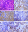
Lymphoplasmacyte-rich meningioma with atypical cystic-solid feature: A case report
- PMID: 33024789
- PMCID: PMC7520769
- DOI: 10.12998/wjcc.v8.i18.4272
Lymphoplasmacyte-rich meningioma with atypical cystic-solid feature: A case report
Abstract
Background: Lymphoplasmacyte-rich meningioma (LPRM) is one of the rarest variants of meningioma and is classified as grade I (benign) tumor. It is characterized by abundant infiltrates of lymphocytes and plasma cells. Here, we report an extremely rare case of LPRM with an atypical imaging finding of multiple cysts around a solid mass.
Case summary: The patient was a 36-year-old man with intermittent headache, dizziness, and vomiting for 2 years. Computed tomography and magnetic resonance imaging presented a cystic solid mass in the right frontal lobe with heavy peritumoral edema and obvious contrast enhancement. The patient was treated with right frontotemporal craniotomy, and gross total resection of the tumor was achieved without adjuvant therapy. There was no clinical or neuroradiological evidence of recurrent or residual tumor for 3 years after initial surgery.
Conclusion: LPRM is one of the rarest variants of meningioma. Although, the mass of this case had common features, multiple cysts with nonuniform size and thin wall around the solid part are uncommon imaging finding, increasing the rate of misdiagnosis. The definitive diagnosis of LPRM relies on histopathological findings.
Keywords: Case report; Computed tomography; Lymphocyte; Lymphoplasmacyte-rich meningioma; Magnetic resonance imaging; Plasmacyte.
©The Author(s) 2020. Published by Baishideng Publishing Group Inc. All rights reserved.
Conflict of interest statement
Conflict-of-interest statement: The authors declare that they have no conflict of interest.
Figures


References
-
- Louis DN, Perry A, Reifenberger G, von Deimling A, Figarella-Branger D, Cavenee WK, Ohgaki H, Wiestler OD, Kleihues P, Ellison DW. The 2016 World Health Organization Classification of Tumors of the Central Nervous System: a summary. Acta Neuropathol. 2016;131:803–820. - PubMed
-
- Liu JL, Zhou JL, Ma YH, Dong C. An analysis of the magnetic resonance imaging and pathology of intracal lymphoplasmacyte-rich meningioma. Eur J Radiol. 2012;81:968–973. - PubMed
-
- Banerjee AK, Blackwood W. A subfrontal tumour with the features of plasmocytoma and meningioma. Acta Neuropathol. 1971;18:84–88. - PubMed
-
- Wiestler OD, Wolf HK. [Revised WHO classification and new developments in diagnosis of central nervous system tumors] Pathologe. 1995;16:245–255. - PubMed
Publication types
LinkOut - more resources
Full Text Sources

