. 2020 Aug 29:19:8-18.
doi: 10.1016/j.omto.2020.08.017. eCollection 2020 Dec 16.

Valeric Acid Suppresses Liver Cancer Development by Acting as a Novel HDAC Inhibitor

Affiliations

Valeric Acid Suppresses Liver Cancer Development by Acting as a Novel HDAC Inhibitor

Rui Han et al. Mol Ther Oncolytics. .

Abstract

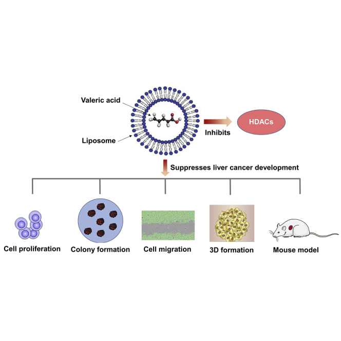
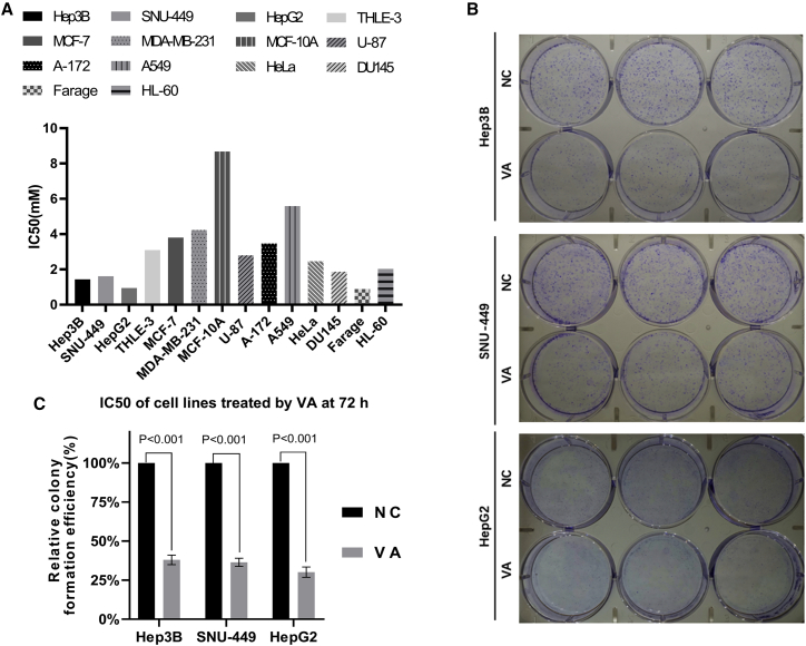
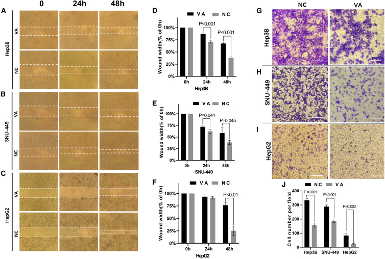
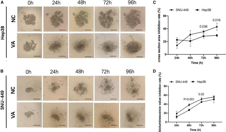
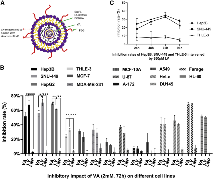
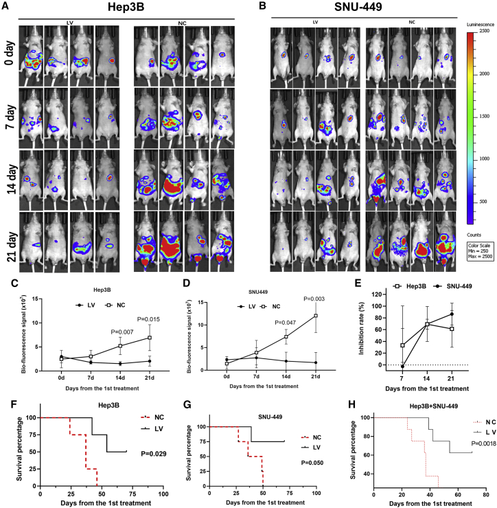
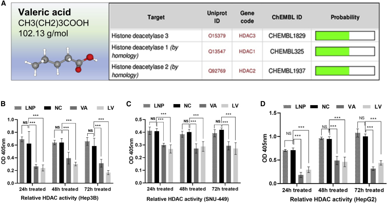
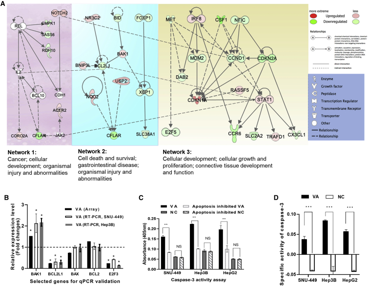
Liver cancer is the fastest growing cause of cancer deaths in the United States due to its aggressiveness and lack of effective therapies. The current preclinical study examines valeric acid (pentanoic acid [C5H10O2]), one of the main compounds of valerian root extract, for its therapeutic use in liver cancer treatment. Anticancer efficacy of valeric acid was tested in a series of in vitro assays and orthotopic xenograft mouse models. The molecular target of valeric acid was also predicted, followed by functional confirmation. Valeric acid has a broad spectrum of anticancer activity with specifically high cytotoxicity for liver cancer in cell proliferation, colony formation, wound healing, cell invasion, and 3D spheroid formation assays. Mouse models further demonstrate that systematic administration of lipid-based nanoparticle-encapsulated valeric acid significantly reduces the tumor burden and improves survival rate. Histone deacetylase (HDAC)-inhibiting functions of valeric acid are also revealed by a structural target prediction tool and HDAC activity assay. Further transcriptional profiling and network analyses illustrate that valeric acid affects several cancer-related pathways that may induce apoptosis. In summary, we demonstrate for the first time that valeric acid suppresses liver cancer development by acting as a potential novel HDAC inhibitor, which warrants further investigation on its therapeutic implications.

Keywords: 3D formation; HDACi; apoptosis; cell migration; cell proliferation; colony formation; hepatocellular carcinoma; lipid nanoparticle; mouse model; valeric acid.


Figures

None
Graphical abstract
Figure 1
Figure 1
Anticancer Effect of Valeric Acid (VA) in Cell Proliferation and Colony Formation Assays (A) The IC50 values were calculated based on dose-response data (cell absorbance) at 72 h. Liver cancer cell lines Hep3B, SNU-449, and HepG2 were sensitive to the treatment of VA. (B) Images from the colony formation assay using Hep3B, SNU-449, and HepG2 cells. (C) The relative colony formation efficiency showed a significant reduction (∼70%) of colonies formed after VA treatment in all 3 liver cancer cell lines tested. Data are presented as the mean ± standard deviation (SD); NS, not significant; NC, negative control.
Figure 2
Figure 2
VA Inhibited Migration and Invasion of Liver Cancer Cells (A–C) Images from the wound healing assay using 3 liver cancer cell lines, Hep3B (A), SNU-449 (B), and HepG2 (C). (D–F) Pictures were taken at 0, 24, and 48 h, and the bar graphs present the percentage of wound recovery in Hep3B (D), SNU-449 (E), and HepG2 (F). The assay was performed in triplicate for each cell line. The wound in the NC groups closed significantly faster than VA groups, respectively. (G–I) Images taken at 24 h from the Transwell invasion assay using liver cancer cell lines Hep3B (G), SNU-449 (H), and HepG2 (I). The average cell number was calculated from counting three randomly chosen different fields. (J) A significantly smaller number of cells was observed in VA-treated groups. Data are presented as the mean ± SD; NS, not significant; NC, negative control.
Figure 3
Figure 3
VA Restrained the 3D Formation Ability of Liver Cancer Cells (A and B) The representative 3D spheroid models of Hep3B (A) and SNU-449 (B) cells treated by VA and NC. The relative 3D formation efficiency calculated from both the cross-section area and fluorescence values showed a significant reduction at all time points (p < 0.01) in VA-treated groups compared to control groups. (C and D) Relative inhibition rates of SNU-449 and Hep3B in response to VA were calculated by comparing the cross-section area (C) and fluorescence values (D) of VA groups to NC at 24, 48, 72, and 96 h, respectively. All assays were performed in triplicate.
Figure 4
Figure 4
LNP Increases Anticancer Efficacy of VA in Liver Cancer (A) A diagram of lipid nanoparticle-encapsulated VA (LV) with double-layer structure composed by PEG bad egg/cholesterol/DODMA. (B) Compared to the VA-treated group, the increased anti-cell proliferation effect was only detected for HCC cell lines Hep3B and SNU-449 but not for hepatoblastoma cell HepG2. (C) Relative inhibition rates were calculated by comparing the OD value of LV to NC at the concentration of 850 μM of LV, which gives over 30% inhibitory rates for liver cancer cells Hep3B and SNU-449 and less than 10% for normal liver cell THLE-3. Data are presented as the mean ± SD; NS, not significant; NC, negative control. ∗p < 0.05; ∗∗p < 0.01;∗∗∗p < 0.001.
Figure 5
Figure 5
LV Suppressed HCC Development and Improved the Survival Rate in the Mouse Study (A and B) The images of bioluminescence of HCC implanted in xenograft mice of Hep3B (A) or SNU-449 (B), from 0 to the 21st day of treatment, were displayed. (C and D) The bioluminescence value was significantly lower in the LV group compared to NC, at 14 and 21 days of treatment, in both Hep3B (C) and SNU-449 (D) cell line mice models. (E) The inhibition rates were calculated by comparing the biofluorescence signal of the LV group to that of NC. (F and G) LV also improved the survival rate of mice implanted with Hep3B (F) or SNU-449 (G) cells compared to NC groups. (H) Combined survival curve of both Hep3B and SNU-449 implanted mice. Data are presented as the mean ± SD; NS, not significant; NC, negative control.
Figure 6
Figure 6
VA Targeted HDAC Enzymes and Reduced Their Activities (A) Three members of HDAC (HDAC1, HDAC2, and HDAC3) were predicted to be the potential targets of VA by the Swiss Target Prediction Tool. (B–D) The relative HDAC activity values in the VA- or LV-treated groups were significantly lower than the activity values in NC at 24, 48, and 72 h in Hep3B (B), SNU-449 (C), and HepG2 (D) cells. No significant differences were observed between the LNP and NC groups. Data are presented as the mean ± SD; NS, not significant; NC, negative control, ∗p < 0.05; ∗∗p < 0.01; ∗∗∗p < 0.001.
Figure 7
Figure 7
Global Transcriptional Impact of VA on Cancer-Related Networks (A) A combined network from genes significantly affected by VA. These networks include “Cancer; cellular development; organismal injury and abnormalities,” Cell death and survival, gastrointestinal disease; organismal injury and abnormalities, and “Cellular development; cellular growth and proliferation; connective tissue development and function.” (B) Consistent expression results from both array and qPCR analyses for 5 selected genes. (C) Significantly increased CASP3 activities were detected in the VA-treated group compared to NC in all cell lines. (D) In SNU-449, Hep3B, and HepG2 cells, VA-treated groups have significantly higher SA values compared to the control groups, respectively (p < 0.001). Values represent the mean ± SD. NS, nonsignificant; NC, negative control. ∗p < 0.05; ∗∗p < 0.01; ∗∗∗p < 0.001.

References

    1. Niino M., Matsuda T. Incidence rates of liver cancer in the world from the Cancer Incidence in Five Continents XI. Jpn. J. Clin. Oncol. 2019;49:693–694. - PubMed
    1. Zhou M., Wang H., Zeng X., Yin P., Zhu J., Chen W., Li X., Wang L., Wang L., Liu Y. Mortality, morbidity, and risk factors in China and its provinces, 1990–2017: a systematic analysis for the Global Burden of Disease Study 2017. Lancet. 2019;394:1145–1158. - PMC - PubMed
    1. Prevention of Infection Related Cancer (PIRCA) Group, Specialized Committee of Cancer Prevention and Control, Chinese Preventive Medicine Association. Non-communicable & Chronic Disease Control and Prevention Society, Chinese Preventive Medicine Association. Health Communication Society, Chinese Preventive Medicine Association [Strategies of primary prevention of liver cancer in China: expert consensus (2018)] Zhonghua Yu Fang Yi Xue Za Zhi. 2019;53:36–44. - PubMed
    1. Hobbs C. Phu: valerian and other anti-hysterics in European and American medicine (1733-1936) Pharm. Hist. 1990;32:132–137. - PubMed
    1. Zare A., Khaksar Z., Sobhani Z., Amini M. Analgesic Effect of Valerian Root and Turnip Extracts. World J. Plast. Surg. 2018;7:345–350. - PMC - PubMed

LinkOut - more resources