

Significant difference between sirolimus and paclitaxel nanoparticles in anti-proliferation effect in normoxia and hypoxia: The basis of better selection of atherosclerosis treatment
- PMID: 33024904
- PMCID: PMC7530254
- DOI: 10.1016/j.bioactmat.2020.09.005
Significant difference between sirolimus and paclitaxel nanoparticles in anti-proliferation effect in normoxia and hypoxia: The basis of better selection of atherosclerosis treatment
Abstract
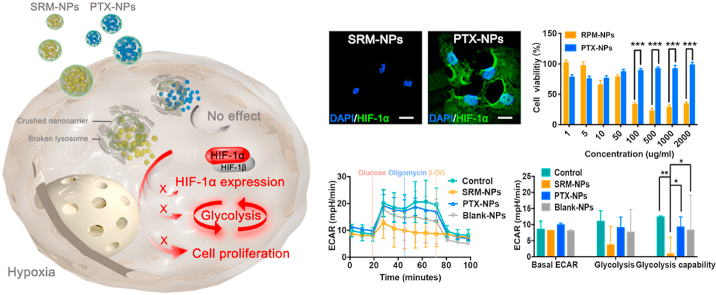
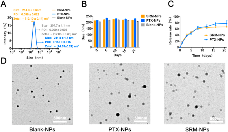
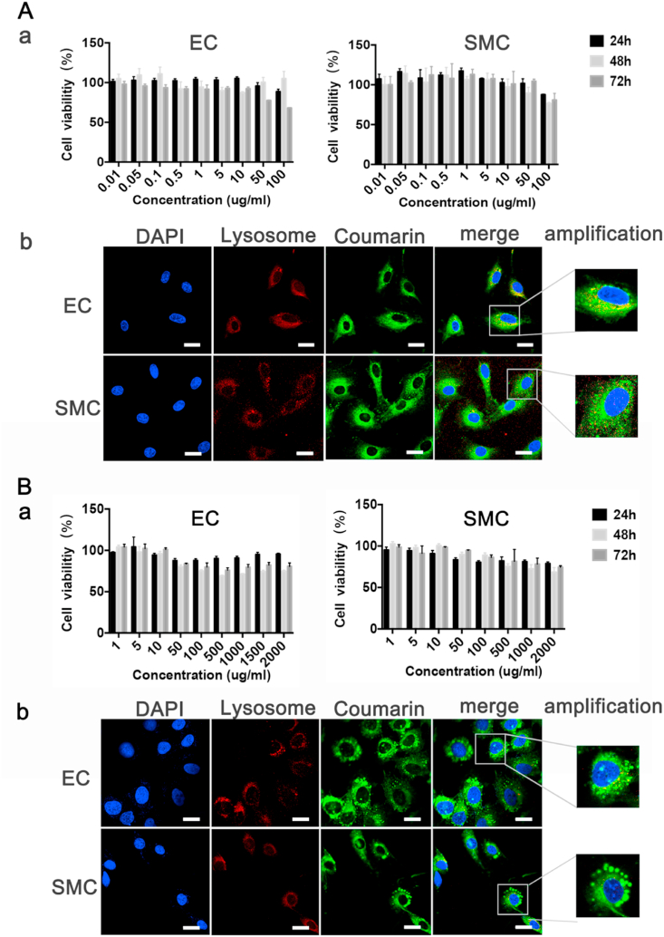
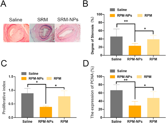
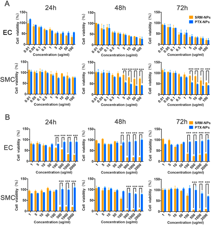
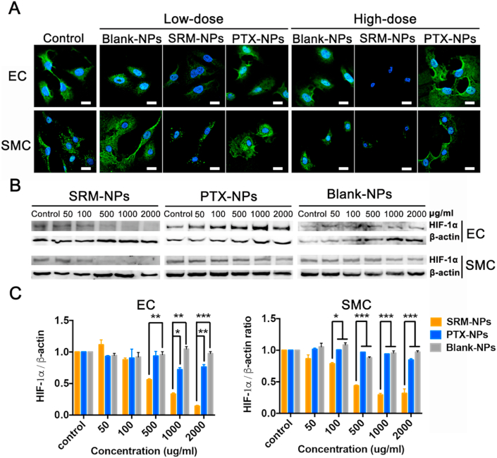
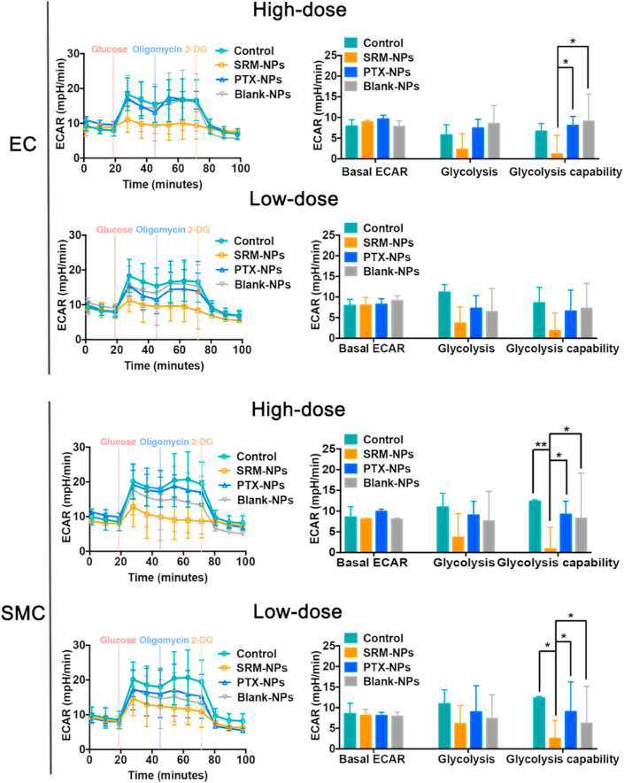
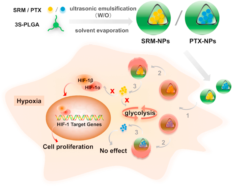
Compared with paclitaxel, sirolimus has been more used in the treatment of vascular restenosis gradually as an anti-proliferative drug, but few basic studies have elucidated its mechanism. The anti-proliferative effects of sirolimus or paclitaxel have been demonstrated by numerous studies under normoxia, but few studies have been achieved focusing hypoxia. In this study, porcine carotid artery injury model and classical cobalt chloride hypoxia cell model were established. Sirolimus nanoparticles (SRM-NPs), paclitaxel nanoparticles (PTX-NPs) and blank nanoparticles (Blank-NPs) were prepared respectively. The effect of RPM-NPs on the degree of stenosis, proliferative index and the expression of PCNA after 28 days of porcine carotid artery injury model was evaluated. Compared with saline group and SRM groups, SRM-NPs group suppressed vascular stenosis, proliferative index and the expression of PCNA (P < 0.01 and P < 0.05). Endothelial cell (EC) and smooth muscle cell (SMC) were pre-treated with cobaltous chloride, followed by SRM-NPs, PTX-NPs, Blank-NPs or PBS control treating, the effects on cell proliferation, HIF-1 expression and glycolysis were detected. SRM-NPs could inhibit EC and SMC proliferation under hypoxia, while PTX-NPs couldn't (P < 0.001). Significant differences between sirolimus and paclitaxel NPs in anti-proliferation effect under normoxia and hypoxia may due to the different inhibitory effects on HIF-1α expression and glycolysis. In conclusion, these results suggest that sirolimus can inhibit the proliferation of hypoxic cells more effectively than paclitaxel. These observations may provide a basis for understanding clinical vascular stenosis therapeutic differences between rapamycin and paclitaxel.
Keywords: Atherosclerosis; Glycolysis; HIF-1α; Hypoxia; Paclitaxel; Sirolimus.
© 2020 [The Author/The Authors].
Conflict of interest statement
The authors declare that they have no known competing financial interests or personal relationships that could have appeared to influence the work reported in this paper.
Figures
Similar articles
-
[Efficacy and mechanism of local delivery of rapamycin and rapamycin-loaded poly(lactic-co-glycolic) acid nanoparticles on coronary restenosis of injury-stenosis model of minipigs].Zhonghua Yi Xue Za Zhi. 2016 Jan 5;96(1):36-42. doi: 10.3760/cma.j.issn.0376-2491.2016.01.009. Zhonghua Yi Xue Za Zhi. 2016. PMID: 26792606 Chinese.
-
Bilayered Nanoparticles with Sequential Release of VEGF Gene and Paclitaxel for Restenosis Inhibition in Atherosclerosis.ACS Appl Mater Interfaces. 2017 Aug 23;9(33):27522-27532. doi: 10.1021/acsami.7b08312. Epub 2017 Aug 9. ACS Appl Mater Interfaces. 2017. PMID: 28748694
-
Sirolimus increases the anti-cancer effect of Huai Er by regulating hypoxia inducible factor-1α-mediated glycolysis in hepatocellular carcinoma.World J Gastroenterol. 2022 Aug 28;28(32):4600-4619. doi: 10.3748/wjg.v28.i32.4600. World J Gastroenterol. 2022. PMID: 36157928 Free PMC article.
-
Drug-eluting stents.Arch Cardiol Mex. 2006 Jul-Sep;76(3):297-319. Arch Cardiol Mex. 2006. PMID: 17091802 Review.
-
How does hypoxia inducible factor-1α participate in enhancing the glycolysis activity in cervical cancer?Ann Diagn Pathol. 2013 Jun;17(3):305-11. doi: 10.1016/j.anndiagpath.2012.12.002. Epub 2013 Feb 1. Ann Diagn Pathol. 2013. PMID: 23375385 Review.
Cited by
-
Benefits and Challenges of Drug-Coated Balloons in Peripheral Artery Disease: From Molecular Mechanisms to Clinical Practice.Int J Mol Sci. 2024 Aug 11;25(16):8749. doi: 10.3390/ijms25168749. Int J Mol Sci. 2024. PMID: 39201436 Free PMC article. Review.
-
PKM2 crotonylation reprograms glycolysis in VSMCs, contributing to phenotypic switching.Oncogene. 2025 Jul;44(24):1990-2003. doi: 10.1038/s41388-025-03353-9. Epub 2025 Apr 3. Oncogene. 2025. PMID: 40181154
-
Top Five Stories of the Cellular Landscape and Therapies of Atherosclerosis: Current Knowledge and Future Perspectives.Curr Med Sci. 2024 Feb;44(1):1-27. doi: 10.1007/s11596-023-2818-2. Epub 2023 Dec 7. Curr Med Sci. 2024. PMID: 38057537 Review.
-
PFKFB3 controls acinar IP3R-mediated Ca2+ overload to regulate acute pancreatitis severity.JCI Insight. 2024 May 23;9(13):e169481. doi: 10.1172/jci.insight.169481. JCI Insight. 2024. PMID: 38781030 Free PMC article.
-
Genetic alterations in coronary cell lines exposed to sirolimus and paclitaxel.Arch Toxicol. 2025 Sep;99(9):3825-3837. doi: 10.1007/s00204-025-04101-4. Epub 2025 Jun 6. Arch Toxicol. 2025. PMID: 40481235
References
-
- Hong T.J., Lee H.W., Choi J.H., Choi J.C., Ahn J., Park J.S. Comparison of long term clinical outcomes between bare metal stent versus different types of drug eluting stents for treatment of acute myocardial infarction. Atherosclerosis. 2017;263:e155–e156.
-
- Mortier P., De Beule M., Carlier G., VanImpe R., Verhegghe B., Verdonck P. Numerical study of the uniformity of balloon-expandable stent deployment. J. Biomech. Eng. 2008;130(2) - PubMed
-
- Nakagawa M., Ohno T., Maruyama R., Okubo M., Nagatsu A., Inoue M. Sesquiterpene lactone suppresses vascular smooth muscle cell proliferation and migration via inhibition of cell cycle progression. Biol. Pharmaceut. Bull. 2007;30(9):1754–1757. - PubMed
LinkOut - more resources
Full Text Sources
Other Literature Sources
Miscellaneous
