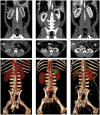
A 30% incidence of renal cysts with varying sizes and densities in biomedical research swine is not associated with renal dysfunction
- PMID: 33024949
- PMCID: PMC7529335
- DOI: 10.1002/ame2.12135
A 30% incidence of renal cysts with varying sizes and densities in biomedical research swine is not associated with renal dysfunction
Abstract
Background: Renal cystic disease arising from various etiologies results in fluid-filled cavities within the kidneys. Moreover, preexisting renal dysfunction has been shown to exacerbate multiple pathologies. While swine bred for biomedical research are often clinically inspected for illness/parasites, more advanced diagnostics may aid in uncovering underlying renal abnormalities.
Methods: Computed tomography was performed in 54 female prepubertal Yorkshire swine to characterize renal cysts; urine and blood chemistry, and histology of cysts were also performed.
Results: Digital reconstruction of right and left kidneys demonstrated that roughly one-third of the animals (17/54; 31%) had one or more renal cyst. Circulating biomarkers of renal function were not different between animals that had cysts and those that did not. Alternatively, urinary glucose (P = .03) was higher and sodium (P = .07) tended to be lower in animals with cysts compared to animals without, with no differences in protein (P = .14) or potassium (P = .20). Aspiration of cystic fluid was feasible in two animals, which revealed that the cystic fluid urea nitrogen (97.6 ± 28.7 vs 911.3 ± 468.2 mg/dL), potassium (29.8 ± 14.4 vs 148.2 ± 24.85 mmol/L), uric acid (2.55 ± 1.35 vs 11.4 ± 5.65 mg/dL), and creatinine (60.34 ± 17.26 vs 268.99 ± 95.79 mg/dL) were much lower than in the urine. Histology demonstrated a cyst that markedly compresses the adjacent cortex and is lined by a single layer of flattened epithelium, bounded by fibrous connective tissue which extends into the parenchyma. There is tubular atrophy and loss in these areas.
Conclusion: This study provides valuable insight for future studies focusing on kidney function in swine bred for biomedical research.
Keywords: computed tomography; cyst; kidney; renal dysfunction; swine.
Published 2020. This article is a U.S. Government work and is in the public domain in the USA. Animal Models and Experimental Medicine published by John Wiley & Sons Australia, Ltd on behalf of The Chinese Association for Laboratory Animal Sciences.
Conflict of interest statement
None.
Figures


Similar articles
-
[Effect of a simple kidney cyst on renal function].Urologiia. 2023 Sep;(4):75-81. Urologiia. 2023. PMID: 37850285 Russian.
-
Hemorrhagic Renal Cyst, a Case Report.J Educ Teach Emerg Med. 2020 Jan 15;5(1):V1-V3. doi: 10.21980/J8C92V. eCollection 2020 Jan. J Educ Teach Emerg Med. 2020. PMID: 37465604 Free PMC article.
-
Renal cysts in living donor kidney transplantation: long-term follow-up in 25 patients.Transplant Proc. 2009 Dec;41(10):4047-51. doi: 10.1016/j.transproceed.2009.09.077. Transplant Proc. 2009. PMID: 20005339
-
Simple renal cyst and renal dysfunction: A pilot study using dimercaptosuccinic acid renal Scan.Nephrology (Carlton). 2016 Aug;21(8):687-92. doi: 10.1111/nep.12654. Nephrology (Carlton). 2016. PMID: 26481869
-
Epithelial transport in polycystic kidney disease.Physiol Rev. 1998 Oct;78(4):1165-91. doi: 10.1152/physrev.1998.78.4.1165. Physiol Rev. 1998. PMID: 9790573 Review.
Cited by
-
Spontaneous Polycystic Kidneys with Chronic Renal Failure in an Aged House Musk Shrew (Suncus murinus).Vet Sci. 2022 Mar 8;9(3):123. doi: 10.3390/vetsci9030123. Vet Sci. 2022. PMID: 35324851 Free PMC article.
-
A case of hydronephrosis due to intrarenal ureteral obstruction in a Japanese Black calf.J Vet Med Sci. 2024 Nov 1;86(11):1162-1167. doi: 10.1292/jvms.24-0173. Epub 2024 Sep 27. J Vet Med Sci. 2024. PMID: 39343540 Free PMC article.
-
Influence of a simple cyst on kidney function.Front Med (Lausanne). 2024 Aug 16;11:1381942. doi: 10.3389/fmed.2024.1381942. eCollection 2024. Front Med (Lausanne). 2024. PMID: 39219799 Free PMC article.
References
-
- Zerres K, Mucher G, Becker J, et al. Prenatal diagnosis of autosomal recessive polycystic kidney disease (ARPKD): molecular genetics, clinical experience, and fetal morphology. Am J Med Genet. 1998;76(2):137‐144. - PubMed
-
- McHugh K, Stringer DA, Hebert D, Babiak CA. Simple renal cysts in children: diagnosis and follow‐up with US. Radiology. 1991;178(2):383‐385. - PubMed
-
- Fick GM, Duley IT, Johnson AM, Strain JD, Manco‐Johnson ML, Gabow PA. The spectrum of autosomal dominant polycystic kidney disease in children. J Am Soc Nephrol. 1994;4(9):1654‐1660. - PubMed
-
- Gabow PA, Chapman AB, Johnson AM, et al. Renal structure and hypertension in autosomal dominant polycystic kidney disease. Kidney Int. 1990;38(6):1177‐1180. - PubMed
-
- Baert L, Steg A. On the pathogenesis of simple renal cysts in the adult. A microdissection study. Urol Res. 1977;5(3):103‐108. - PubMed
LinkOut - more resources
Full Text Sources

