[Preprint]. 2020 Sep 29:2020.09.28.317685.
doi: 10.1101/2020.09.28.317685.

SARS-CoV-2 D614G Variant Exhibits Enhanced Replication ex vivo and Earlier Transmission in vivo

Affiliations

SARS-CoV-2 D614G Variant Exhibits Enhanced Replication ex vivo and Earlier Transmission in vivo

Yixuan J Hou et al. bioRxiv. .

Update in

  • SARS-CoV-2 D614G variant exhibits efficient replication ex vivo and transmission in vivo.
    Hou YJ, Chiba S, Halfmann P, Ehre C, Kuroda M, Dinnon KH 3rd, Leist SR, Schäfer A, Nakajima N, Takahashi K, Lee RE, Mascenik TM, Graham R, Edwards CE, Tse LV, Okuda K, Markmann AJ, Bartelt L, de Silva A, Margolis DM, Boucher RC, Randell SH, Suzuki T, Gralinski LE, Kawaoka Y, Baric RS. Hou YJ, et al. Science. 2020 Dec 18;370(6523):1464-1468. doi: 10.1126/science.abe8499. Epub 2020 Nov 12. Science. 2020. PMID: 33184236 Free PMC article.

Abstract

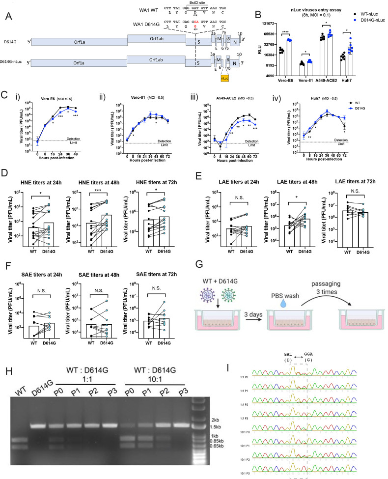
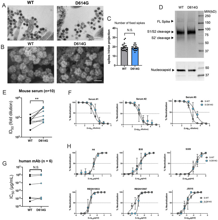
The D614G substitution in the S protein is most prevalent SARS-CoV-2 strain circulating globally, but its effects in viral pathogenesis and transmission remain unclear. We engineered SARS-CoV-2 variants harboring the D614G substitution with or without nanoluciferase. The D614G variant replicates more efficiency in primary human proximal airway epithelial cells and is more fit than wildtype (WT) virus in competition studies. With similar morphology to the WT virion, the D614G virus is also more sensitive to SARS-CoV-2 neutralizing antibodies. Infection of human ACE2 transgenic mice and Syrian hamsters with the WT or D614G viruses produced similar titers in respiratory tissue and pulmonary disease. However, the D614G variant exhibited significantly faster droplet transmission between hamsters than the WT virus, early after infection. Our study demonstrated the SARS-CoV2 D614G substitution enhances infectivity, replication fitness, and early transmission.


Figures

Figure 1.
Figure 1.. SARS-CoV-2 D614G variant demonstrate enhanced infectivity in some cell lines and replication fitness in upper respiratory epithelia.
A. Genomes of recombinant SARS-CoV-2 D614G variants. B. Entry efficiency of WT-nLuc and D614G-nLuc in multiple cell lines at MOI of 0.1. After 1h infection, cells were cultured in the medium containing neutralization antibodies to minimize the secondary round of infection. The relative light unit (RLU) representing the nLuc expression level was measured at 8h post infection. C. Multi-step growth curves of the two variants at Vero-E6 (i), Vero-81 (ii) and A549-ACE2 (iii) and Huh7 (iv) cell lines at MOI = 0.5. Comparison of 24, 48 and 72h titers between the two variants infected primary nasal (D), large airway (E) and small airway (F) cells in triplicate. Triplicated titers of the two viruses in the cultures form the same donor were analyzed by paired t-test. G. Schematic of competition assay on large airway epithelial cells. Cultures were infected with 1:1 or 10:1 ratio of WT and D614G mixture at MOI at 0.5, and serially passaged three times. H. BstCI digestion of the partial S gene from the competition assay samples. A 1.5kb fragment containing the residue 614 was amplified from the total RNA collected from competition assay. I. Sanger sequencing chromatogram of S RNA collected from the competition assay. Data between the WT and D614G viruses in B and C are analyzed using unpaired t-test, and the data between the two groups in D, E and F are analyzed using paired t-test. N.S., not significantly different, p > 0.05; *, p < 0.05 **, p < 0.01; ***, p < 0.001.
Figure 2.
Figure 2.. D614G substitution does not alter SARS-CoV-2 virion morphology and S protein cleavage pattern but change viral sensitivity to neutralizing antibodies.
A. Transmission electron microscopy image of WT and D614G virions on airway epithelial cell surface, scale bar: 200 nm. B. Scanning electron microscopy images of WT and D614G virions on airway epithelial cell surface, scale bar: 100 nm. C. Quantification of S protein on individual SARS-CoV-2 virion projections. The number of S protein on individual virion projections from different SEM images were quantify manually, n=20. D. Western blot analysis of SARS-CoV-2 virions washed from WT- or D614G-infected LAE culture surface at 72h. Each lane contains mixed sample from triplicated cultures. Full-length (S), S1/S2 cleaved and S2’ cleaved spike protein (upper panel) and nucleocapsid protein (lower panel) were probed. E. ID50 values of 10 serum samples collected from D614-form Spike-vaccinated mice neutralizing WT- and D614G-nLuc viruses. F. Three representative neutralization curves of the mouse sera against both viruses. Summarized IC50 values (G) and individual neutralization curves (H) of 6 human nAbs against both viruses. Data between the WT and D614G viruses in E and G are analyzed using paired t-test. N.S., not significantly different, p > 0.05; **, p < 0.01.
Figure 3.
Figure 3.. D614G variant exhibit similar pathogenesis but faster transmission than the WT virus in vivo.
A. Lung, brain and nasal turbinate titers of WT and D614G infected hACE2 mice were determined on day 2 (i) day 5 (ii). Each mouse was infected with 105 PFU of the virus, n= 5/group, plaque assay detection limit (1.7 log10PFU/mL) is indicated in a dash line. Viral titers of lung (B) and nasal turbinates (C) collected from SARS-CoV-2 infected hamsters at day 3 and 6. Each hamster was infected with 103 PFU of virus, plaque assay detection limit (1 log10PFU/mL) is indicated in a dash line. D. Body weight of mock-, WT- and D614G-infected hamsters, n = 4/group. E. Immunohistochemistry (IHC) staining of SARS-CoV-2 nucleocapsid protein in the representative lung tissues collected from WT- and D614G-infected hamsters, scale bar = 100 μm. F. H&E staining of representative lung tissues collected on day 3, 6, and 9 from hamsters infected with WT or D614G, scale bar : 1mm. G-i: Quantification of IHC positive cells in hamster lung tissues, following scoring system: 0, no positive cell; 1, <10%; 2, 10–50%; 3, >50% of positive cells in each lobe of lung. G-ii. The size of pulmonary lesion was determined based on the mean percentage of affected area in each section of the collected lobes from each animal. G-iii Pathological severity scores in infected hamsters, based on the percentage of inflammation area for each section of the five lobes collected from each animal following scoring system: 0, no pathological change; 1, affected area (≤10%); 2, affected area (<50%, >10%); 3, affected area (≥50%); an additional point was added when pulmonary edema and/or alveolar hemorrhage was observed. (H) Viral titers in nasal washes collected from the infected and exposed hamster pairs in WT and D614G groups; plaque assay detection limit (1 log10 PFU/mL) is indicated in a dash line. Data between the WT and D614G viruses in A, B, C, D, and G are analyzed using unpaired t-test. The number of transmitted hamsters at different timepoints are; analyzed by Fisher exact test. N.S., not significantly different, p >0.05.

Similar articles

Cited by

References

    1. Wichmann D., Sperhake J.-P., Lütgehetmann M., Steurer S., Edler C., Heinemann A., Heinrich F., Mushumba H., Kniep I., Schröder A. S., Burdelski C., de Heer G., Nierhaus A., Frings D., Pfefferle S., Becker H., Bredereke-Wiedling H., de Weerth A., Paschen H.-R., Sheikhzadeh-Eggers S., Stang A., Schmiedel S., Bokemeyer C., Addo M. M., Aepfelbacher M., Püschel K., Kluge S., Autopsy Findings and Venous Thromboembolism in Patients With COVID-19. Annals of Internal Medicine. 173, 268–277 (2020). - PMC - PubMed
    1. Cheung E. W., Zachariah P., Gorelik M., Boneparth A., Kernie S. G., Orange J. S., Milner J. D., Multisystem Inflammatory Syndrome Related to COVID-19 in Previously Healthy Children and Adolescents in New York City. JAMA. 324, 294–296 (2020). - PMC - PubMed
    1. Mao R., Liang J., Shen J., Ghosh S., Zhu L.-R., Yang H., Wu K.-C., Chen M.-H., Implications of COVID-19 for patients with pre-existing digestive diseases. The Lancet Gastroenterology & Hepatology. 5, 425–427 (2020). - PMC - PubMed
    1. Hou Y. J., Okuda K., Edwards C. E., Martinez D. R., Asakura T., Dinnon K. H., Kato T., Lee R. E., Yount B. L., Mascenik T. M., Chen G., Olivier K. N., Ghio A., Tse L. V., Leist S. R., Gralinski L. E., Schäfer A., Dang H., Gilmore R., Nakano S., Sun L., Fulcher M. L., Livraghi-Butrico A., Nicely N. I., Cameron M., Cameron C., Kelvin D. J., de Silva A., Margolis D. M., Markmann A., Bartelt L., Zumwalt R., Martinez F. J., Salvatore S. P., Borczuk A., Tata P. R., Sontake V., Kimple A., Jaspers I., O’Neal W. K., Randell S. H., Boucher R. C., Baric R. S., SARS-CoV-2 Reverse Genetics Reveals a Variable Infection Gradient in the Respiratory Tract. Cell. 182, 429–446.e14 (2020). - PMC - PubMed
    1. Korber B., Fischer W. M., Gnanakaran S., Yoon H., Theiler J., Abfalterer W., Hengartner N., Giorgi E. E., Bhattacharya T., Foley B., Hastie K. M., Parker M. D., Partridge D. G., Evans C. M., Freeman T. M., de Silva T. I., Angyal A., Brown R. L., Carrilero L., Green L. R., Groves D. C., Johnson K. J., Keeley A. J., Lindsey B. B., Parsons P. J., Raza M., Rowland-Jones S., Smith N., Tucker R. M., Wang D., Wyles M. D., McDanal C., Perez L. G., Tang H., Moon-Walker A., Whelan S. P., LaBranche C. C., Saphire E. O., Montefiori D. C., Tracking Changes in SARS-CoV-2 Spike: Evidence that D614G Increases Infectivity of the COVID-19 Virus. Cell. 182, 812–827.e19 (2020). - PMC - PubMed

Publication types