[Preprint]. 2020 Sep 29:2020.09.22.20199125.
doi: 10.1101/2020.09.22.20199125.

Molecular Architecture of Early Dissemination and Massive Second Wave of the SARS-CoV-2 Virus in a Major Metropolitan Area

Affiliations

Molecular Architecture of Early Dissemination and Massive Second Wave of the SARS-CoV-2 Virus in a Major Metropolitan Area

S Wesley Long et al. medRxiv. .

Update in

Abstract

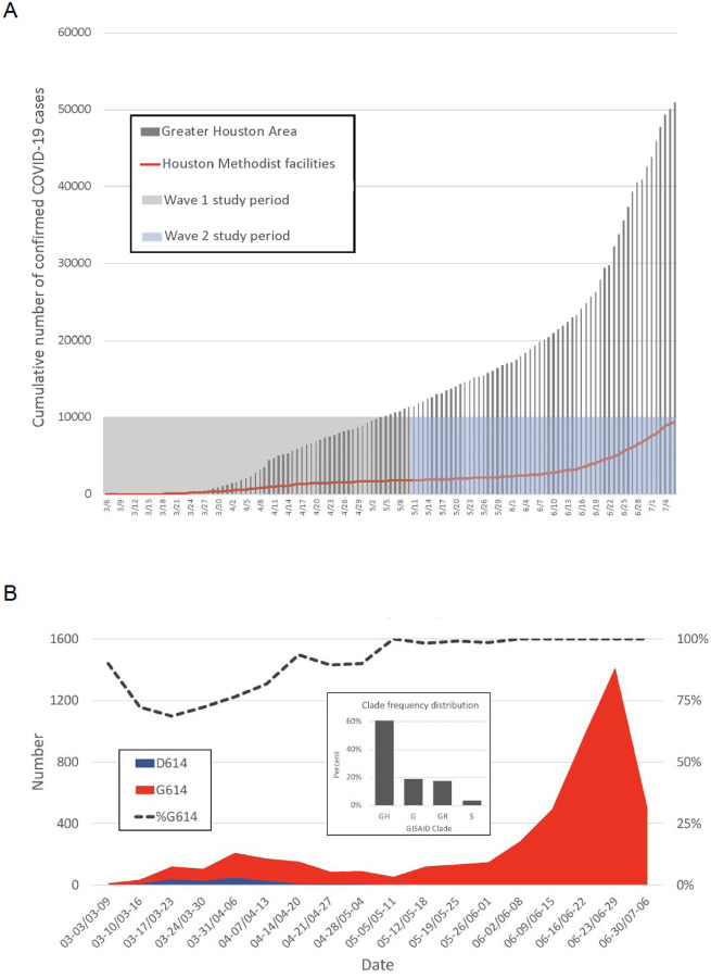
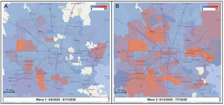
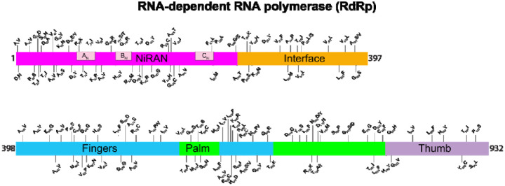
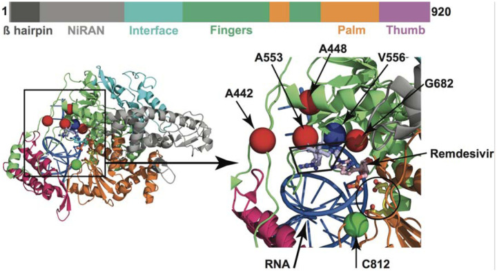
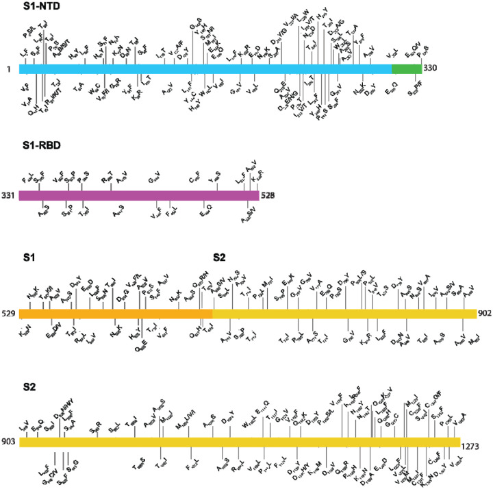
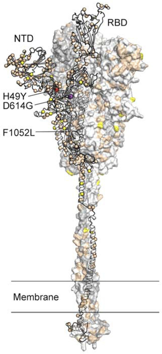
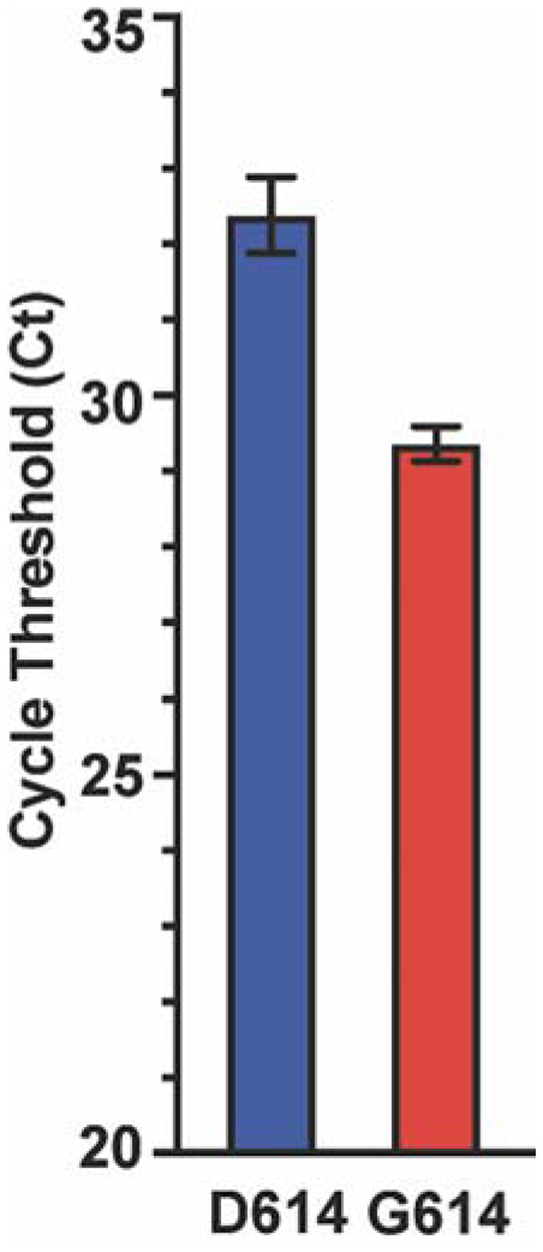
We sequenced the genomes of 5,085 SARS-CoV-2 strains causing two COVID-19 disease waves in metropolitan Houston, Texas, an ethnically diverse region with seven million residents. The genomes were from viruses recovered in the earliest recognized phase of the pandemic in Houston, and an ongoing massive second wave of infections. The virus was originally introduced into Houston many times independently. Virtually all strains in the second wave have a Gly614 amino acid replacement in the spike protein, a polymorphism that has been linked to increased transmission and infectivity. Patients infected with the Gly614 variant strains had significantly higher virus loads in the nasopharynx on initial diagnosis. We found little evidence of a significant relationship between virus genotypes and altered virulence, stressing the linkage between disease severity, underlying medical conditions, and host genetics. Some regions of the spike protein - the primary target of global vaccine efforts - are replete with amino acid replacements, perhaps indicating the action of selection. We exploited the genomic data to generate defined single amino acid replacements in the receptor binding domain of spike protein that, importantly, produced decreased recognition by the neutralizing monoclonal antibody CR30022. Our study is the first analysis of the molecular architecture of SARS-CoV-2 in two infection waves in a major metropolitan region. The findings will help us to understand the origin, composition, and trajectory of future infection waves, and the potential effect of the host immune response and therapeutic maneuvers on SARS-CoV-2 evolution.


Figures

FIG 1
FIG 1
(A) Confirmed COVID-19 cases in the Greater Houston Metropolitan region. Cumulative number of COVID-19 patients over time through July 7, 2020. Counties include Austin, Brazoria, Chambers, Fort Bend, Galveston, Harris, Liberty, Montgomery, and Waller. The shaded area represents the time period during which virus genomes characterized in this study were recovered from COVID-19 patients. The red line represents the number of COVID-19 patients diagnosed in the Houston Methodist Hospital Molecular Diagnostic Laboratory. (B) Distribution of strains with either the Asp614 or Gly614 amino acid variant in spike protein among the two waves of COVID-19 patients diagnosed in the Houston Methodist Hospital Molecular Diagnostic Laboratory. The large inset shows major clade frequency for the time frame studied.
FIG 2
FIG 2
Sequential time-series heatmaps for all COVID-19 Houston Methodist patients during the study period. Geospatial distribution of COVID-19 patients is based on zip code. Panel A (left) shows geospatial distribution of sequenced SARS-CoV-2 strains in wave 1 and panel B (right) shows wave 2 distribution. The collection dates are shown at the bottom of each panel. The insets refer to numbers of strains in the color spectrum used. Note difference in numbers of strains used in panel A and panel B insets.
FIG 3
FIG 3
Location of amino acid replacements in RNA-dependent RNA polymerase (RdRp/Nsp12) among the 5,085 genomes of SARS-CoV-2 sequenced. The various RdRp domains are color-coded. The numbers refer to amino acid site. Note that several amino acid sites have multiple variants identified.
FIG 4
FIG 4
Amino acid changes identified in Nsp12 (RdRp) in this study that may influence interaction with remdesivir. The schematic at the top shows the domain architecture of Nsp12. (Left) Ribbon representation of the crystal structure of Nsp12-remdesivir monophosphate-RNA complex (PDB code: 7BV2). The structure in the right panel shows a magnified view of the boxed area in the left panel. The Nsp12 domains are colored as in the schematic at the top. The catalytic site in Nsp12 is marked by a black circle in the right panel. The side chains of amino acids comprising the catalytic site of RdRp (Ser758, Asp759, and Asp760) are shown as balls and stick and colored yellow. The nucleotide binding site is boxed in the right panel. The side chains of amino acids participating in nucleotide binding (Lys544, Arg552, and Arg554) are shown as balls and sticks and colored light blue. Remdesivir molecule incorporated into the nascent RNA is shown as balls and sticks and colored light pink. The RNA is shown as a blue cartoon and bases are shown as sticks. The positions of Cα atoms of amino acids identified in this study are shown as red and green spheres and labeled. The amino acids that are shown as red spheres are located above the nucleotide binding site, whereas Cys812 located at the catalytic site is shown as a green sphere. The side chain of active site residue Ser758 is shown as ball and sticks and colored yellow. The location of Cα atoms of remdesivir resistance conferring amino acid Val556 is shown as blue sphere and labeled.
FIG 5
FIG 5
Location of amino acid replacements in spike protein among the 5,085 genomes of SARS-CoV-2 sequenced. The various spike protein domains are color-coded. The numbers refer to amino acid site. Note that many amino acid sites have multiple variants identified.
FIG 6
FIG 6
Location of amino acid substitutions mapped on the SARS-CoV-2 spike protein. Model of the SARS-CoV-2 spike protein with one protomer shown as ribbons and the other two protomers shown as a molecular surface. The Cα atom of residues found to be substituted in one or more virus isolates identified in this study is shown as a sphere on the ribbon representation. Residues found to be substituted in 1–9 isolates are colored tan, 10–99 isolates yellow, 100–999 isolates colored red (H49Y and F1052L), and >1000 isolates purple (D614G). The surface of the aminoterminal domain (NTD) that is distal to the trimeric axis has a high density of substituted residues. RBD, receptor binding domain.
FIG 7
FIG 7
Cycle threshold (Ct) for every SARS-CoV-2 patient sample tested using the Hologic Panther assay. Data are presented as mean +/− standard error of the mean for strains with an aspartate (D614, n=102 strains, blue) or glycine (G614, n=812 strains, red) at amino acid 614 of the spike protein. Mann-Whitney test, *P<0.0001.
FIG 8
FIG 8
Biochemical characterization of spike RBD variants. (A) Size-exclusion chromatography (SEC) traces of the indicated spike-RBD variants. Dashed line indicates the elution peak of spike-6P. (B) The relative expression of all RBD variants as determined by the area under the SEC traces. All expression levels are normalized relative to spike-6P. (C) Thermostability analysis of RBD variants by differential scanning fluorimetry. Each sample had three replicates and only mean values were plotted. Black vertical dashed line indicates the first melting temperature of 6P-D614G and orange vertical dashed line indicates the first melting temperature of the least stable variant (spike-G446V). (D) First apparent melting temperature of all RBD variants. (E) ELISA-based binding affinities for ACE2 and (F) the neutralizing antibody CR3022 to the indicated RBD variants. (G) Summary of EC50s for all measured RBD variants.

Similar articles

Cited by

References

    1. 2020. World Health Organization Coronavirus Disease 2019 (COVID-19) Situation Report. https://www.who.int/docs/default-source/coronaviruse/situation-reports/2.... Accessed April 21.
    1. Gorbalenya AE, Baker SC, Baric RS, de Groot RJ, Drosten C, Gulyaeva AA, Haagmans BL, Lauber C, Leontovich AM, Neuman BW, Penzar D, Perlman S, Poon LLM, Samborskiy DV, Sidorov IA, Sola I, Ziebuhr J, Coronaviridae Study Group of the International Committee on Taxonomy of V. 2020. The species Severe acute respiratory syndrome-related coronavirus: classifying 2019-nCoV and naming it SARS-CoV-2. Nature Microbiology 5:536–544. - PMC - PubMed
    1. Wang C, Horby PW, Hayden FG, Gao GF. 2020. A novel coronavirus outbreak of global health concern. Lancet 395:470–473. - PMC - PubMed
    1. Perlman S. 2020. Another Decade, Another Coronavirus. New England Journal of Medicine 382:760–762. - PMC - PubMed
    1. Allel K, Tapia-Muñoz T, Morris W. 2020. Country-level factors associated with the early spread of COVID-19 cases at 5, 10 and 15 days since the onset. Glob Public Health doi:10.1080/17441692.2020.1814835:1-14 - DOI - PubMed

Publication types