Review
. 2020 Nov;125(11):1124-1134.
doi: 10.1007/s11547-020-01279-8. Epub 2020 Oct 6.

Myocarditis: imaging up to date

Affiliations
Review

Myocarditis: imaging up to date

Carlo Liguori et al. Radiol Med. 2020 Nov.

Abstract

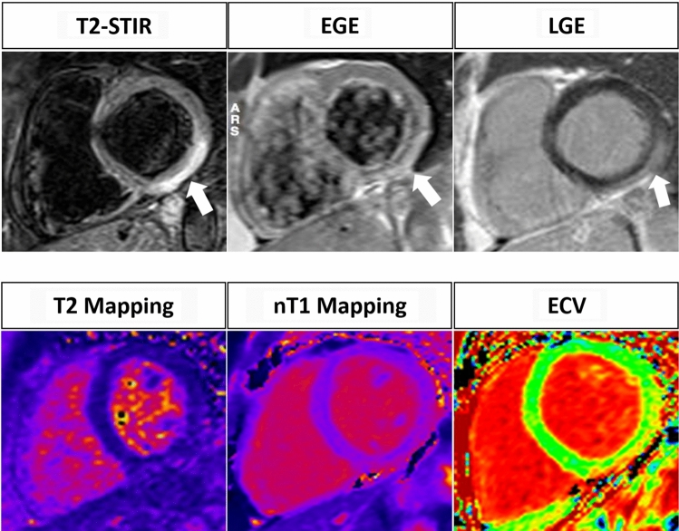
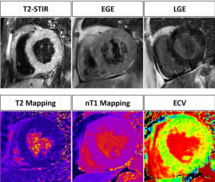
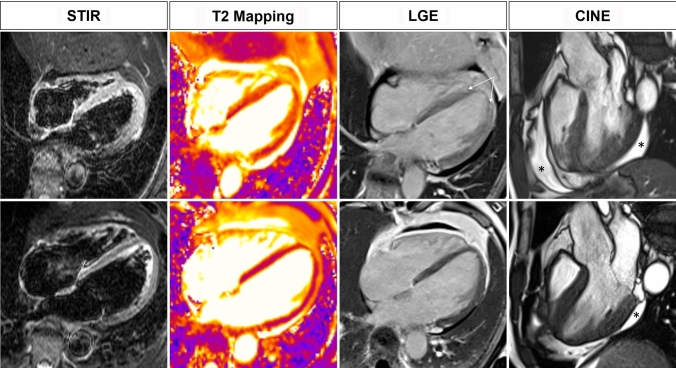
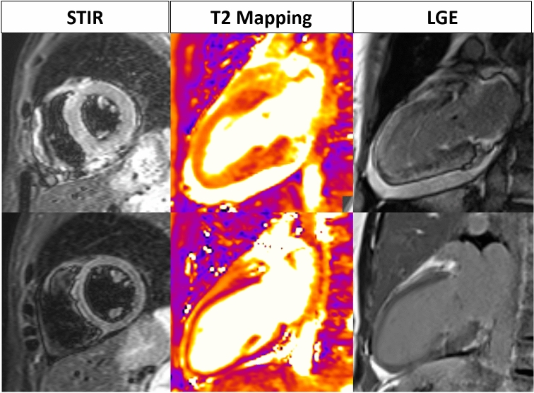
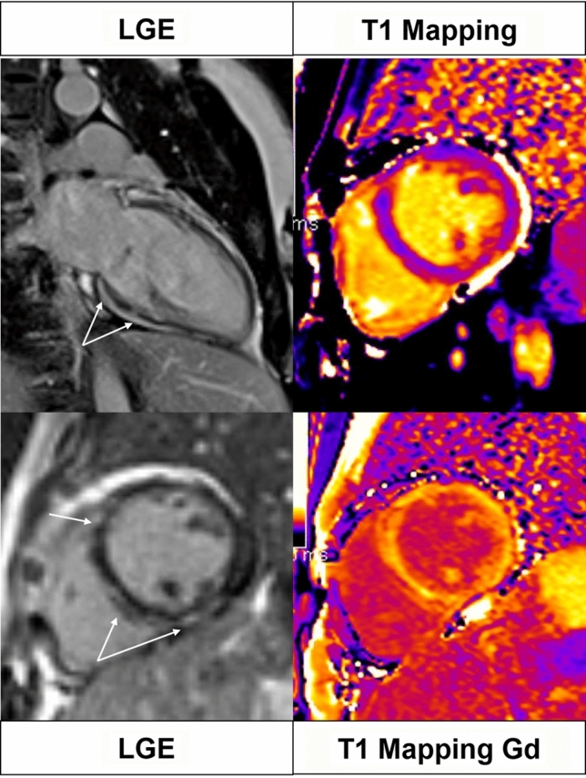
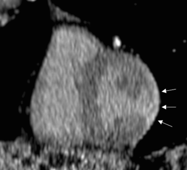
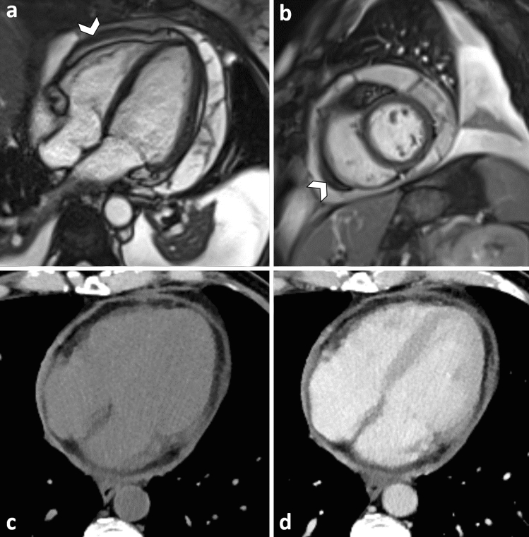
Myocarditis is an inflammatory disease of the heart muscle, diagnosed by histological, immunological, and immunohistochemical criteria. Endomyocardial biopsy represents the diagnostic gold standard for its diagnosis but is infrequently used. Due to its noninvasive ability to detect the presence of myocardial edema, hyperemia and necrosis/fibrosis, Cardiac MR imaging is routinely used in the clinical practice for the diagnosis of acute myocarditis. Recently pixel-wise mapping of T1 and T2 relaxation time have been introduced into the clinical Cardiac MR protocol increasing its accuracy. Our paper will review the role of MR imaging in the diagnosis of acute myocarditis.

Keywords: Cardiac MR; Magnetic resonance imaging; Myocardial inflammation; Myocarditis.


Conflict of interest statement

The authors have nothing to disclose.

Figures

Fig. 1
Fig. 1
A 57 year old female with sudden onset of retrosternal chest pain. T2-w STIR image shows an hyperintense subepicardial rim representing myocardial edema in the inferior wall of the LV (arrow). EGE image shows an increase of Gd uptake in the same area (arrow). On LGE image an hyperintense area confirming the presence of myocardial necrosis can be observed in the inferior wall of the LV (arrow). The diagnosis of myocarditis can be performed with old LLC (3 criteria out of 3). Mapping images confirm the findings of acute myocardial inflammation: T2 mapping value is higher than 60 ms; nT1 value is higher than 1100 ms and ECV is higher than 32%. Revised LLC are also positive for acute myocardial inflammation (2 criteria out of 2)
Fig. 2
Fig. 2
A 55 year old male with malaise and an EF of 33%. No edema, hyperemia and necrosis can be observed in T2-w, EGE and LGE images, respectively. Mapping sequences show an overall value of 58 ms on T2 mapping, an overall value of 1150 ms on nT1 mapping and an overall ECV value of 35%. The diagnosis of acute myocarditis cannot be obtained with the original LLC (0 criteria out of 3), but is provided by applying the revised LLC (2 criteria out of 2)
Fig. 3
Fig. 3
43 year old male with chest pain, and depressed systolic function (20%) during sepsis by Pseudomonas aeruginosa. CMR scan performed in the acute phase (upper row) shows hypersignal of the septum and mid-apical lateral wall, mirrored by prolonged T2 on mapping; these findings are consistent with edema. PSIR shows patchy areas of subepicardial enhancement in the apical segments (arrows). Pericardial effusion (asterisk). Early follow-up scan obtained 30 days later shows normalization of T2 and regression of the enhancement of the myocardium; near complete resolution of the pericardial effusion
Fig. 4
Fig. 4
COVID-19 myocarditis in a 54-year old female. In the acute phase (upper row), STIR and T2 mapping depict global edema, resulting in swelling of the myocardium. LGE shows diffuse enhancement with subepicardial gradient. Clinical findings and EF (43%) suggested acute fulminant form. On a follow-up scan (lower row) performed 55 days later near complete regression of radiological findings is documented
Fig. 5
Fig. 5
38-year old female with polimorphic extrasystole, regressing on effort, and mild mitral valve regurgitation. Diffuse areas of myocardial enhancement are seen on basal and midcavity inferoseptal and anteroseptal wall, with subepicardial and midwall distribution (arrows). The same segments display focal anomalies of both native and post-contrast T1 mapping. Findings are consistent with chronic myocarditis
Fig. 6
Fig. 6
CT examination performed in a patient with chest pain. Late phase acquisition (8 min after contrast administration) using low Kv. Short-axis reformation shows subepicardial enhancement in the mid-ventricular lateral wall, suggestive of myocarditis. The diagnosis was later confirmed by lab test and MRI scan
Fig. 7
Fig. 7
Cine MRI scan on 4-chamber (a) and short-axis (b) view, shows circumferential pericardial effusion with multiple linear septa within the pericardial sac, indicating fibrin deposition. Note thickening of the visceral pericardium (arrowheads). CT before (c) and after contrast (d) shows diffuse thickening and enhancement of the pericardium in a patient affected by lung carcinoma

Similar articles

Cited by

References

    1. Richardson P, McKenna W, Bristow M, et al. Report of the 1995 World Health Organization/International Society and Federation of Cardiology Task Force on the definition and classification of cardiomyopathies. Circulation. 1996;93:841–842. doi: 10.1161/01.CIR.93.5.841. - DOI - PubMed
    1. Leone O, Veinot JP, Angelini A, et al. Consensus statement on endomyocardial biopsy from the Association for European Cardiovascular Pathology and the Society for Cardiovascular Pathology. Cardiovasc Pathol. 2012;21:245–274. doi: 10.1016/j.carpath.2011.10.001. - DOI - PubMed
    1. Kindermann I, Barth C, Mahfoud F, et al. Update on myocarditis. J Am Coll Cardiol. 2012;59:779–792. doi: 10.1016/j.jacc.2011.09.074. - DOI - PubMed
    1. Monney P, Locca D, Muzzarelli S, et al. Cardiac magnetic resonance in acute myocarditis: A new non-invasive diagnostic gold standard? Rev Med Suisse. 2012;8(343):1177–1183. - PubMed
    1. Caforio AL, Pankuweit S, Arbustini E, et al. Current state of knowledge on aetiology, diagnosis, management, and therapy of a position statement of the European Society of Cardiology Working Group on Myocardial and Pericardial Diseases. Eur Heart J. 2013;34:2636–2648d. doi: 10.1093/eurheartj/eht210. - DOI - PubMed

MeSH terms