

Erosion of a right ventricular pacer lead into the left chest wall
- PMID: 33025306
- PMCID: PMC7538471
- DOI: 10.1186/s40792-020-00999-3
Erosion of a right ventricular pacer lead into the left chest wall
Abstract
Background: Erosion of a pacer lead into the chest wall may result in pericardial effusion with cardiac tamponade. Free rupture into the pleura or mediastinum can result in hypotension and cardiac arrest.
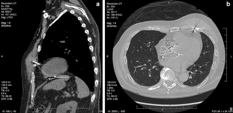
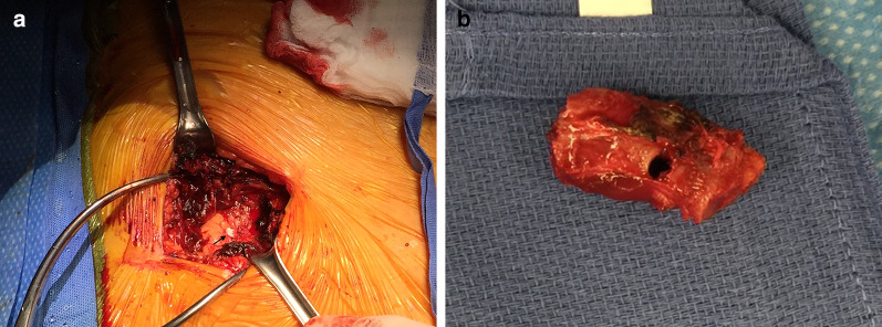
Case presentation: We report a unique case of a right ventricular pacer lead which eroded through the right ventricle into the left chest wall and penetrated a rib. The patient presented with a tender chest wall mass without pericardial or pleural effusion. The segment of rib which the pacing lead had penetrated was removed.
Conclusions: The patient tolerated the procedure well and was discharged 1 week after the operation. This case adds to the current literature the justification of removal of temporary and non-functional pacing leads.
Keywords: Chest wall; Pacing lead; Rib perforation.
Conflict of interest statement
HEG receives patent royalties/licensing fees Garrett, Bromet for sternal closure device not used or described in this report. MJH, JMC, and DSW have no competing interests.
Figures
References
-
- Satpathy R, Hee T, Esterbrooks D, Mohiuddin S. Delayed defibrillator lead perforation: an increasing phenomenon. Pacing Clin Electrophysiol. 2008;31(1):10–12. - PubMed
LinkOut - more resources
Full Text Sources
