Review
. 2020 Oct 5;21(19):7357.
doi: 10.3390/ijms21197357.

Cellular and Molecular Mechanisms of CD8+ T Cell Differentiation, Dysfunction and Exhaustion

Affiliations
Review

Cellular and Molecular Mechanisms of CD8+ T Cell Differentiation, Dysfunction and Exhaustion

Daniel J Verdon et al. Int J Mol Sci. .

Abstract

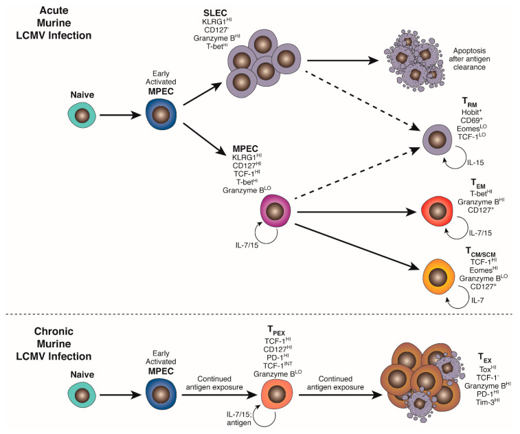
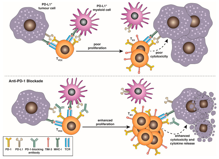
T cells follow a triphasic distinct pathway of activation, proliferation and differentiation before becoming functionally and phenotypically "exhausted" in settings of chronic infection, autoimmunity and in cancer. Exhausted T cells progressively lose canonical effector functions, exhibit altered transcriptional networks and epigenetic signatures and gain constitutive expression of a broad coinhibitory receptor suite. This review outlines recent advances in our understanding of exhausted T cell biology and examines cellular and molecular mechanisms by which a state of dysfunction or exhaustion is established, and mechanisms by which exhausted T cells may still contribute to pathogen or tumour control. Further, this review describes our understanding of exhausted T cell heterogeneity and outlines the mechanisms by which checkpoint blockade differentially engages exhausted T cell subsets to overcome exhaustion and recover T cell function.

Keywords: PD-1; T cell exhaustion; cancer; chronic viral infections; epigenetics; immunotherapy; inhibitory receptors.


Conflict of interest statement

The authors declare that they have no conflict of interest.

Figures

Figure 1
Figure 1
Acute and chronic infection drive distinct programs of CD8+ T cell differentiation. Activated naïve CD8+ cells initiate a program of metabolic, transcriptional and epigenetic changes that facilitate differentiation into KLRG1HI CD127neg effector and KLRG1neg CD127HI memory precursors (MPEC). In an acute infection, an expanded pool of terminally differentiated cytotoxic effectors (SLEC/TEFF) clear infected cells and subsequently contract, leaving behind a heterogeneous pool of MPEC-derived self-renewing stem-like (TSCM) and central memory (TCM) in secondary lymphoid organs and effector memory (TEM) and resident memory (TRM) in peripheral tissues to provide protection against secondary exposure to the same pathogen. Chronic TCR stimulation, exacerbated by the absence of appropriate CD4+ T cell “help” and co-stimulatory and cytokine signalling, drives an alternative program of differentiation and epigenetic remodeling mediated by NFATc1, BATF, IRF4 and Tox. This gives rise to a TCF-1+PD-1INT pool of self-renewing “precursor” exhausted cells carrying a distinct epigenetic signature (TPEX) that produce and continually replenish an expanded pool of terminally exhausted progeny (TEX) able to partially control, but not fully clear, established infection. TEX maintain the epigenetic signature established in TPEX and exhibit a spectrum of effector function impairment and high expression of a suite of co-inhibitory receptors.
Figure 2
Figure 2
TPEX respond to anti-PD-1/PD-L1 blockade. TCF-1HI intratumoral or virus-specific precursor-exhausted T cells (TPEX) expressing intermediate surface levels of PD-1 continually replenish and maintain a pool of cytotoxic granzyme BHI TCF-1NEG PD-1HI TIM-3HI exhausted progeny (TEX) when they are stimulated. PD-1:PD-L1 interactions impair activation and proliferation of TPEX and activation cytokine production and direct cytotoxicity of TEX. Blockade of these interactions using monoclonal antibodies allows the expansion of a larger tumour- or virus-reactive TEX pool, and enhances direct cytotoxicity against transformed or virus-infected cells.

References

    1. Chen L., Flies D.B. Molecular mechanisms of T cell co-stimulation and co-inhibition. Nat. Rev. Immunol. 2013;13:227–242. doi: 10.1038/nri3405. - DOI - PMC - PubMed
    1. Henry C.J., Ornelles D.A., Mitchell L.M., Brzoza-Lewis K.L., Hiltbold E.M. IL-12 Produced by Dendritic Cells Augments CD8+ T cell Activation through the Production of the Chemokines CCL1 and CCL171. J. Immunol. 2008;181:8576–8584. doi: 10.4049/jimmunol.181.12.8576. - DOI - PMC - PubMed
    1. Díaz-Montero C.M., El-Naggar S.A., Al Khami A., El Naggar R., Montero A.J., Cole D.J., Salem M.L. Priming of naive CD8+ T cells in the presence of IL-12 selectively enhances the survival of CD8+CD62Lhi cells and results in superior anti-tumor activity in a tolerogenic murine model. Cancer Immunol. Immunother. 2007;57:563–572. doi: 10.1007/s00262-007-0394-0. - DOI - PMC - PubMed
    1. Ramos H.J., Davis A.M., Cole A.G., Schatzle J.D., Forman J., Farrar J.D. Reciprocal responsiveness to interleukin-12 and interferon-α specifies human CD8+ effector versus central memory T-cell fates. Blood. 2009;113:5516–5525. doi: 10.1182/blood-2008-11-188458. - DOI - PMC - PubMed
    1. Redeker A., Welten S.P., Baert M.R.M., Vloemans S.A., Tiemessen M.M., Staal F.J.T., Arens R. The Quantity of Autocrine IL-2 Governs the Expansion Potential of CD8+ T Cells. J. Immunol. 2015;195:4792–4801. doi: 10.4049/jimmunol.1501083. - DOI - PubMed

MeSH terms

Substances