

In Vivo Lymphatic Circulating Tumor Cells and Progression of Metastatic Disease
- PMID: 33028044
- PMCID: PMC7650582
- DOI: 10.3390/cancers12102866
In Vivo Lymphatic Circulating Tumor Cells and Progression of Metastatic Disease
Abstract
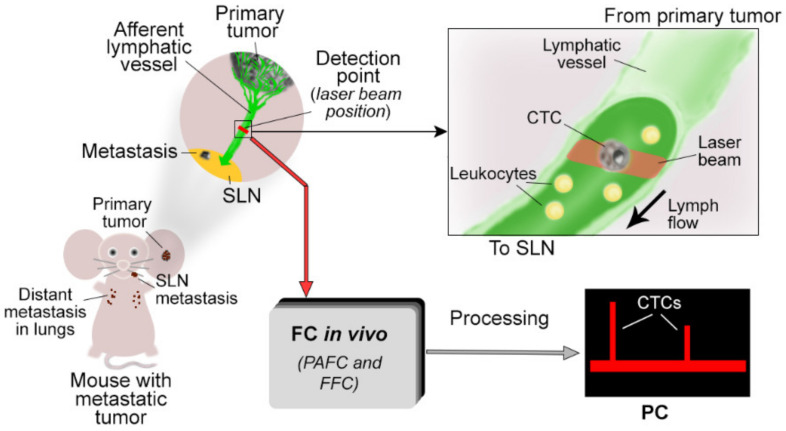
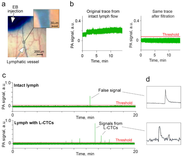
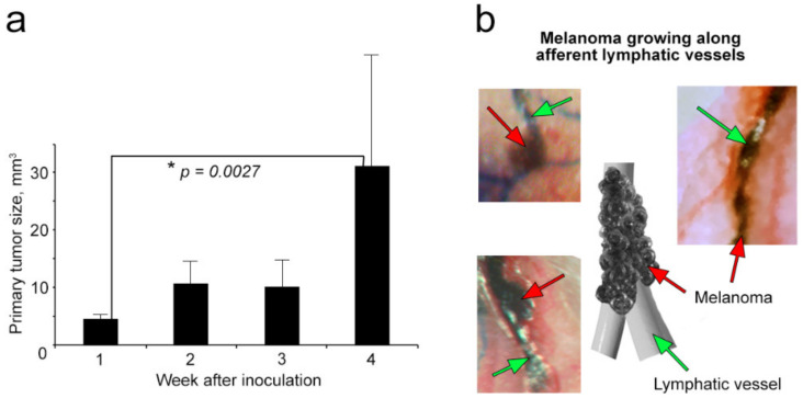
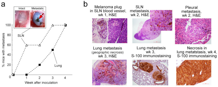
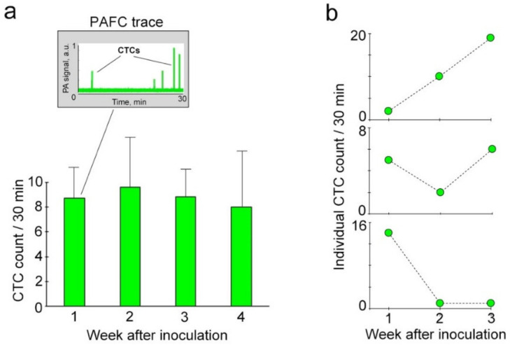
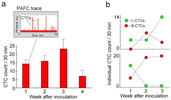
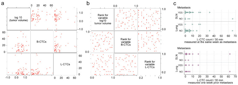
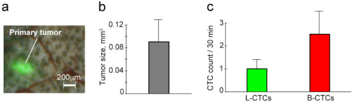
The dissemination of circulating tumor cells (CTCs) by lymph fluid is one of the key events in the development of tumor metastasis. However, little progress has been made in studying lymphatic CTCs (L-CTCs). Here, we demonstrate the detection of L-CTCs in preclinical mouse models of melanoma and breast cancer using in vivo high-sensitivity photoacoustic and fluorescent flow cytometry. We discovered that L-CTCs are be detected in pre-metastatic disease stage. The smallest primary tumor that shed L-CTCs was measured as 0.094mm×0.094mm, its volume was calculated as 0.0004 mm3; and its productivity was estimated as 1 L-CTC per 30 minutes. As the disease progressed, primary tumors continued releasing L-CTCs with certain individual dynamics. The integrated assessment of lymph and blood underlined the parallel dissemination of CTCs at all disease stages. However, the analysis of links between L-CTC counts, blood CTC (B-CTC) counts, primary tumor size and metastasis did not reveal statistically significant correlations, likely due to L-CTC heterogeneity. Altogether, our results showed the feasibility of our diagnostic platform using photoacoustic flow cytometry for preclinical L-CTC research with translational potential. Our findings also demonstrated new insights into lymphatic system involvement in CTC dissemination. They help to lay the scientific foundation for the consideration of L-CTCs as prognostic markers of metastasis and to emphasize the integrative assessment of lymph and blood.
Keywords: circulating tumor cells; in vivo flow cytometry; lymphatic vessels; lymphography; marker; metastasis; personalized prognosis; photoacoustics; sentinel lymph node.
Conflict of interest statement
Vladimir Zharov, Ekaterina Galanzha, and UAMS have a financial interest in the technology discussed in this publication. Vladimir Zharov has a financial interest in CytoAstra, LLC, which has licensed the technology. These financial interests have been reviewed and approved in accordance with the UAMS conflict of interest policies.
Figures
Similar articles
-
Lymph Liquid Biopsy for Detection of Cancer Stem Cells.Cytometry A. 2021 May;99(5):496-502. doi: 10.1002/cyto.a.24221. Epub 2020 Sep 18. Cytometry A. 2021. PMID: 32869909 Free PMC article.
-
Dynamic Fluctuation of Circulating Tumor Cells during Cancer Progression.Cancers (Basel). 2014 Jan 15;6(1):128-42. doi: 10.3390/cancers6010128. Cancers (Basel). 2014. PMID: 24434542 Free PMC article.
-
Mechanical Conditioning (MeCo) Score Progressively Increases Through the Metastatic Cascade in Breast Cancer via Circulating Tumor Cells.Cancers (Basel). 2025 May 12;17(10):1632. doi: 10.3390/cancers17101632. Cancers (Basel). 2025. PMID: 40427129 Free PMC article.
-
Relevance of CTC Clusters in Breast Cancer Metastasis.Adv Exp Med Biol. 2020;1220:93-115. doi: 10.1007/978-3-030-35805-1_7. Adv Exp Med Biol. 2020. PMID: 32304082 Review.
-
Gauging the Impact of Cancer Treatment Modalities on Circulating Tumor Cells (CTCs).Cancers (Basel). 2020 Mar 21;12(3):743. doi: 10.3390/cancers12030743. Cancers (Basel). 2020. PMID: 32245166 Free PMC article. Review.
Cited by
-
Advances in circulating tumor cells for early detection, prognosis and metastasis reduction in lung cancer.Front Oncol. 2024 Jun 21;14:1411731. doi: 10.3389/fonc.2024.1411731. eCollection 2024. Front Oncol. 2024. PMID: 38974237 Free PMC article. Review.
-
Dormancy of cutaneous melanoma.Cancer Cell Int. 2024 Feb 28;24(1):88. doi: 10.1186/s12935-024-03278-5. Cancer Cell Int. 2024. PMID: 38419052 Free PMC article. Review.
-
Targeting circulating tumor cells to prevent metastases.Hum Cell. 2024 Jan;37(1):101-120. doi: 10.1007/s13577-023-00992-6. Epub 2023 Oct 24. Hum Cell. 2024. PMID: 37874534 Free PMC article. Review.
-
Molecular Mechanisms of Lymphatic Metastasis in Breast Cancer: An Updated Review.Cancers (Basel). 2025 Jun 25;17(13):2134. doi: 10.3390/cancers17132134. Cancers (Basel). 2025. PMID: 40647432 Free PMC article. Review.
-
Predictive Factors for Sentinel Lymph Node Positivity in Melanoma Patients-The Role of Liquid Biopsy, MicroRNA and Gene Expression Profile Panels.Cancers (Basel). 2025 Apr 10;17(8):1281. doi: 10.3390/cancers17081281. Cancers (Basel). 2025. PMID: 40282456 Free PMC article. Review.
References
-
- Cristofanilli M., Budd G.T., Ellis M.J., Stopeck A., Matera J., Miller M.C., Reuben J.M., Doyle G.V., Allard W.J., Terstappen L.W.M.M., et al. Circulating tumor cells, disease progression, and survival in metastatic breast cancer. N. Engl. J. Med. 2004;351:781–791. doi: 10.1056/NEJMoa040766. - DOI - PubMed
Grants and funding
LinkOut - more resources
Full Text Sources
Research Materials
